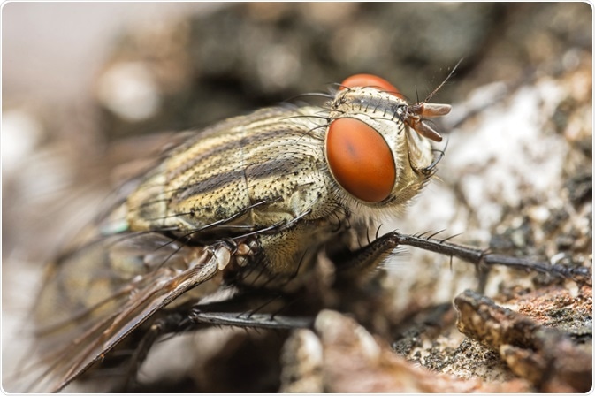
Studying the decomposition of corpses through examining the temperature or the rigidness can be challenging when the body was found by investigators after 48 hours postmortem.

Image Credit: Jan H Andersen/Shutterstock.com
Luckily, forensic entomologists have discovered ways of how insect activity can be used as a means of navigating the location of death and estimating the post-mortem interval of a decomposing body after the 48 hours time range.
The morphological study of early forms of flies can be studied by light microscopy and scanning electron microscopy (SEM) to aid forensic investigations in estimating PMI of a body.
Developmental stages of anthropoids can reveal where and when the death occurred
Forensic entomology aids crime scene investigations through studying insects and arthropods. In some death cases, only eggs and the larva are found on the body.
Insects are known to approach a dead body and oviposition from flies can be found on the body when hours after death have passed. Through analyzing the eggs and the early stages of insects, the conditions of murder such as the post-mortem interval estimation, whether the body had been displaced, the link of a suspect to the crime scene, and the presence of drugs can be determined.
Anthropoids undergo a developmental stage, known as an instar, to reach to the level of sexual maturity from the different stages of molting. The unique stages between instars can be examined from different physical characteristics, such as patterns, head width, colors, and body sizes.
Furthermore, these developmental stages are influenced by the environmental conditions and therefore the surroundings of where the death may have happened can be identified.
The development stages for insects while staying on the corpse can also be studied to calculate the time of death by counting backward from the most recent state of development state. To illustrate, fly eggs are known to hatch under 18 hours with the first instar taking 20 hours to grow, therefore the earliest time that the eggs could have been laid would be 38 hours ago, taking into account the molting of the larvae.
The role of microscopy in forensic entomology
Examining the morphological changes occurring within a fly requires microscopical techniques due to their tiny sizes. More than one microscope is used by forensic entomologists, as each microscope is unique in their ability to study the different characteristics of the eggs and/or larvae found on a corpse.

Image Credit: Rachata Kietsirikul/Shutterstock.com
Light microscopy reveals information on the early stages of the fly life cycle
Light microscopy is known for its feature to study the early stages of Diptera species, examining their organs such as spines or spiracles of greater specimens as well as the mouthparts, serving as internal structures of the species. It further provides quick results through a simpler technique than other microscopes, which plays importance in solving forensic cases that require fast processing of the specimen.
The internal organ in the fly larvae, cephalopharingeal skeleton, can be examined with light microscopy without the need of dissecting the body wall, as there are no movements in the position for all sclerites.
However, light microscopy may be limited in providing descriptions of small structures in some developmental stages as well as tiny specimens of eggs and instars I and II. The technique may only become advantageous in distinguishing species that are entirely different in morphology and describing instar III with pupae.
As a result, providing details in between the early and later stages of larval stages may also give out challenges when not bred in a laboratory. The resolution of light microscopy may also be insufficient in capturing the details of some structures other than the external larvae morphology in all instars.
Using scanning electron microscopy to identify the morphology of body larval
This type of microscopy is advantageous for its detailed description in the morphology of developmental stages. Unlike light microscopy, SEM can provide more details on various morphological characteristics to differentiate the species examined without the need for breeding in the laboratory environment.
It is also known to capture detailed structures of instars I and II, some detailed observations that cannot be achieved with the resolution from light microscopy alone. The high resolution of SEM is capable of giving details in specific larval features for identification, such as the respiratory slits of posterior spiracles, the peritrema ornamentation, and the formation of the tegument.
Although this technique gives more details on the larval body morphology to their tiny internal structures, SEM is a more expensive technique than light microscopy and it requires sample preparation before moving forward with the analysis.
To solve this particular issue, Ubero-Pascal, et al. combines both light microscope and SEM to give out the most optimum identification of the larval body morphology. Light microscopy is used for quick specimen identification and to find the relationship between the immature stages. While SEM can be useful to analyze the larval body morphology, from eggs, instars I and II, as well as refining the identification of instar III and pupae from light microscopy.
References:
- Méndez-Vilas, A. and Díaz, J., 2010. Microscopy. 4th ed. Badajoz: Formatex Research Center, pp.1548-1556. https://www.researchgate.net/publication/256250031_Microscopy_and_forensic_entomology
- Mendonça, P., dos Santos-Mallet, J., de Mello, R., Gomes, L., and Queiroz, M. (2008). Identification of fly eggs using scanning electron microscopy for forensic investigations. Micron, 39(7), pp.802-807. https://www.sciencedirect.com/science/article/pii/S0968432808000231?casa_token=lVoEJT4KCqUAAAAA:-C2Mtfo91kbcbrTpcJyL4Mj_F-06a1u6icQvP7Ohz-muOuYSf-F4YTtNS0_1-KQ0EGg3fLIM6pY
- The Australian Museum. 2020. Decomposition - Forensic Evidence. [Online] Available at https://australian.museum/about/history/exhibitions/death-the-last-taboo/decomposition-forensic-evidence/ (Accessed 17 July 2020).
Further Reading