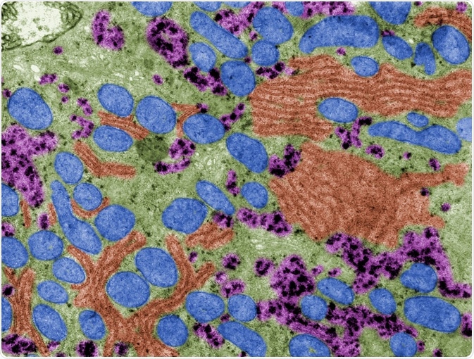
Transmission electron microscopy (TEM) is a powerful tool that provides precise information on the morphology of particles in the nanometer range, thus supporting its continued utility in the emergent diagnosis of viruses.

TEM. Image Credit: Jose Luis Calvo/Shutterstock.com
Early use of TEM for virus discovery
Both Max Knoll and Ernst Ruska constructed the world’s first transmission electron microscope (TEM) during the early 1930s. Helmut Ruska, the younger brother of Ernst Ruska, harnessed this novel microscope to characterize the morphology of serval viruses, as well as determine the size and shape of various viral particles.
Ruska’s discovery of the utility of TEM in clinical virology quickly led to the adoption of this technique for diagnosing both smallpox and chickenpox, which are caused by the variola and varicella-zoster viruses, respectively. These early studies found the varicella-zoster virus, which is within the herpes family of viruses, to have a spherical shape and a diameter within the range of 140 to 150 nanometers (nm).
The utility of negative staining
By 1959, Sydney Brenner and Robert Horne developed a negative staining method for its use in high-resolution electron microscopy like TEM. In negative staining, aqueous-based suspensions of biological particle samples are first deposited onto carbon-coated grids.
Heavy metal salts such as uranyl acetate or phosphotungstic acid are then used to stain the grids to enhance the visibility of the biological particles while also examining their unique structural characteristics. When incorporated into TEM, negative staining not only allowed researchers to distinguish viral particles from the background, but also improved the ability to determine the symmetry of the particles and whether the virus was surrounded by an envelope.
The combined analytical technique of negative staining with TEM allowed for its clinical use throughout both the 1970s and 1980s. In fact, during this time, TEM was successfully used to isolate and identify numerous viruses that continue to have significant clinical impacts on patients, which include adenoviruses, enteroviruses, paramyxoviruses, rotaviruses, and reoviruses.
Despite its utility during this time, TEM was unable to detect other types of important viruses such as hepatitis and those outside of rotaviruses that were responsible for gastroenteritis due to their inability to be cultured in vitro.
Declining use of TEM
TEM is a highly time-consuming technique, as it requires the biological samples to be first embedded into a resin before sectioning with an ultramicrotome. Furthermore, the analysis of TEM images can be particularly laborious and therefore requires a significant amount of knowledge by the individual on viral morphology. Despite these disadvantages, its utility in providing an accurate diagnosis maintained its clinical use until the 1990s.
By the 1990s, several novel molecular techniques including enzyme-linked immunosorbent assays (ELISAs) and polymerase chain reactions (PCRs) were developed and proved to offer greater sensitivity capabilities while also expanding the number of samples that could be tested simultaneously.
As technology continued to advance, several other techniques have also emerged as alternatives to negative staining TEM including immunofluorescence microscopy, endpoint dilution assay (TCID50), and analytical flow cytometry. Taken together, these more sensitive molecular techniques quickly replaced TEM in many aspects of virological diagnosis, particularly in gastroenteritis cases.
Current role of TEM in viral detection and diagnosis
Although several other methods are available for the diagnosis of viruses, TEM remains very useful within medical virology. During the 2003 severe acute respiratory syndrome (SARS) pandemic in Honk Kong and Southern China, for example, TEM was used to characterize the specific etiology of the coronavirus responsible for this outbreak.
In the same year, TEM was used on the other side of the world in the United States to identify a poxvirus known as monkeypox virus that was causing widespread illness to plague prairie dogs.
In fact, as recently as 2020, a combination of both scanning electron microscopy (SEM) and TEM was used to produce early images of the novel severe acute respiratory syndrome coronavirus 2 (SARS-CoV-2). The images produced by these two electron microscopy techniques determined that the SARS-CoV-2 closely resembled both the 2013 SARS virus molecule and the Middle East respiratory syndrome coronavirus (MERS-CoV) that emerged in 2012.
Conclusion
While the role of TEM has transitioned from being routinely used to diagnose viral infections, it remains an important tool in the field of medical virology. In fact, TEM can be used to confirm a diagnosis that was previously established with molecular techniques. TEM also offers the unique ability to simultaneously detect multiple infections that are caused by one or more viruses, which could otherwise be missed in conventional molecular or antigen tests.
Federal agencies like the United States Food and Drug Administration (FDA) and the European Medicines Agency (EMEA) continue to recommend the use of TEM as a complementary tool for various in vitro assays and molecular tests. These agencies specify the use of TEM to assess the viral safety of biopharmaceutical products including cell lines, culture supernatants, and fermenter bulk harvests. Taken together, the “catch-all” properties of TEM make it an important tool in the detection of viruses or virus-like particles that might be present within these materials.
Sources:
- Roingeard, P., Raynal, P., Eymieux, S., & Blanchard, E. (2019). Virus detection by transmission electron microscopy: Still useful for diagnosis and a plus for biosafety. Reviews in Medical Virology 29(1). doi:10.1002/rmv.2019.
- Xiao, C., Chen, X., Xie, Q., et al. (2021). Virus identification in electron microscopy images by residual mixed attention network. Computer Methods and Programs in Biomedicine 198. doi:10.1016/j.cmpb.2020.105766.
- Blancett, C. D., Fetterer, D. P., Koistinen, K. A., et al. (2017). Accurate virus quantitation using a Scanning Transmission Electron Microscopy (STEM) detector in a scanning electron microscope. Journal of Virological Methods 248; 136-144. doi:10.1016.j.jviromet.2017.06.014.
- New Images of Novel Coronavirus SARS-CoV-2 Now Available [Online]. Available from: www.niaid.nih.gov/news-events/novel-coronavirus-sarscov2-images
Further Reading