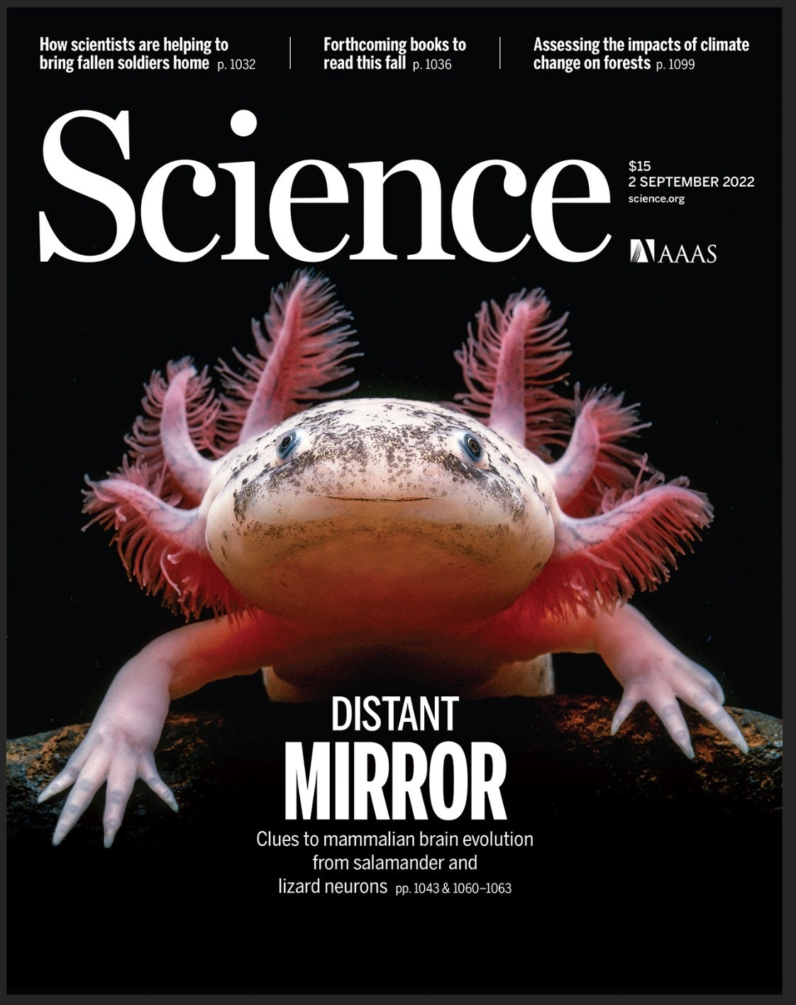
The first spatiotemporal cellular atlas of axolotl (Ambystoma mexicanum) brain growth and regeneration, created by a multi-institute research team under the direction of BGI-Research, reveals how brain damage may recover itself. The study was released as the cover story for the most recent edition of Science.
 Single-cell Stereo-seq reveals induced progenitor cells involved in axolotl brain regeneration published on Science. Image Credit: BGI Group
The study team looked at how the salamander brain develops and regenerates, identified the crucial neural stem cell subsets involved, and detailed how these stem cell subsets rebuild injured neurons. The scientists also discovered that brain regeneration and development have some characteristics, which aid in the development of the cognitive brain and provide new paths for regenerative medicine research and treatment of the nervous system.
Some vertebrates, unlike mammals, have the capacity to regenerate a variety of organs, including a portion of the central nervous system. The axolotl is one of them and has the ability to regenerate the brain in addition to other organs including the limbs, tail, eyes, skin, and liver. Axolotls have a more mammalian-like brain structure than other teleosts like zebrafish because they have undergone more evolution.
As a result, the axolotl was chosen for this study as the perfect model organism for studying brain regeneration.
The cells and pathways that are involved in brain regeneration have only been partially identified by prior studies. In this work, scientists employed BGI’s Stereo-seq technology to produce a single-cell resolution spatiotemporal map of salamander brain development spanning six key developmental phases. This map reveals the chemical properties of different neurons and dynamic changes in spatial distribution.
The ventricular zone region’s neural stem cell subtypes are hard to discern in the early stages of development, but they start to specialize with geographical regional features in the teen stage, according to research. This finding shows that various subtypes may perform various tasks during regeneration.
The investigators were able to examine cell regeneration by taking brain samples at seven different intervals (2, 5, 10, 15, 20, 30, and 60 days) after causing damage to the salamander brain’s cortical region.
On day 15, incomplete tissue connections started to emerge in the wounded area. New neural stem cell subtypes started to develop in the wound location in the early stages of injury. Researchers noticed that the wound had grown new tissues on days 20 and 30, but that the cell makeup was significantly different from that of the unharmed area. The cell types and dispersion had recovered to their pre-injury state by day 60.
Scientists found that the creation process of neurons is very comparable throughout both development and regeneration by analyzing the chemical changes during salamander brain growth and regeneration. This finding suggests that brain damage may cause neural stem cells to alter their normal developmental course and begin the process of regenerating.
Using axolotl as a model organism, we have identified key cell types in the process of brain regeneration. This discovery will provide new ideas and guidance for regenerative medicine in the mammalian nervous system.”
Dr Ying Gu, Study Joint Corresponding Author and Deputy Director, BGI-Research
Dr Ying Gu adds, “The brain is a complex organ with interconnected neurons. Therefore, a major goal in central nervous system regenerative medicine is not only to reconstruct the spatial structure of neurons, but also to reconstruct the specific patterns of their intra-tissue connections. Therefore, it is important to reconstruct the 3D structure of the brain and understand the systemic reactions between brain regions during regeneration in future research.”
Source:
Journal reference:
Wei, X., et al. (2022) Single-cell Stereo-seq reveals induced progenitor cells involved in axolotl brain regeneration. Science. doi.org/10.1126/science.abp9444