At two separate 60× magnifications, the same Yeast sample was collected. Both achieved the same visual contrast. However, with 60X/1.4 NA oil immersion objective, users can obtain 60% shorter illumination times, brighter images and enhanced resolution.
Users can now achieve such pictures in totally autonomous plate scanning with IDEA Bio-Medical’s new precision oil dispensing device on Hermes imaging system.
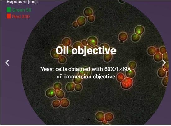
Oil objective. Yeast cells obtained with 60×/1.4 NA oil immersion objective. Image Credit: IDEA Bio-Medical Ltd.
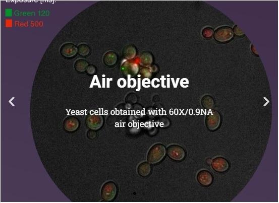
Air objective. Yeast cells obtained with 60×/0.9 NA air objective. Image Credit: IDEA Bio-Medical Ltd.
Overview
Why use high NA oil objectives in high content screening?
- Brighter Images
- Higher Resolution
- Shorter Exposure
Oil immersion objectives in automated microscopy? — Now users can
Oil immersion objectives had hitherto been off-limits for automated microscopy, particularly for high-content screening, due to the need for human involvement to add oil while scanning a sample. Furthermore, when automatically scanning across the sample, the proper quantity of oil must stay precisely between the objective and the sample.
Finally, the matching of the index of refraction, which improves picture resolution and brightness, makes autofocusing more difficult.
IDEA Bio-Medical is aware of the difficulties with automated microscopy, as well as the potential benefits for the life sciences research community. They have developed a unique and automated system that accurately supplies immersion oil just when it is needed, based on their decades of expertise in several domains of ODM and OEM engineering.
Such precision prevents excess oil from collecting and dripping from the bottom of a sample, as well as a lack of oil during a scan.
The autofocus mechanism works perfectly with immersion oil, allowing for crisp and reliable pictures. Cleaning is a snap, thanks to the quick-snap objective exchange technology, which makes removing objectives from the microscope a breeze.
For the first time, a high-content imaging system can automate high-resolution cell biology applications using oil immersion objectives.
Unsupervised and without human involvement, samples in a multi-well plate format can be scanned in their entirety. For live-cell tests, the system can support a range of multi-well plates and sample types, as well as provide environmental control.
Completely automated super-resolution imaging is also possible with high NA oil objectives.
Full automation, no compromises
- No user intervention; no oil spilling
- For easy maintenance, autofocus, X, Y, and Z movements have been optimized, as well as long-duration oil capsules
- Unique hardware automatically adds immersion oil to objectives
- Autonomous, rapid image acquisition for:
- Time-lapse imaging of live cells
- Full-plate scanning
Applications
- Microbiology, Virology and Yeast studies
- Super-resolution radial fluctuations (SRRF) live-cell imaging
- Mitochondria, Endoplasmic Reticulum, Cytoskeleton and Focal Adhesion imaging
- Spot / Foci / Granule visualization
- Fluorescence In-Situ Hybridization (FISH)