Due to their optical transparency and genetic tractability, zebrafish (Danio rerio) are an excellent model organism for the analysis of human disease pathology. The lower cost, higher throughput, and fewer ethical considerations make them a perfect alternative to mammalian screening.
Since there is a wide variety of organs and traits that should be detected, automated zebrafish analysis provides special challenges.
IDEA Bio-Medical is pleased to showcase its one-of-a-kind dedicated imaging platform for automated data gathering and analysis in zebrafish larvae to measure fluorescence, morphological changes, and other parameters in a high-throughput format.
- Rapid collection of multicolor, perfectly focused photos of zebrafish embryos
- For structural co-analysis, combine with fluorescent labeling
- The quickest and most straightforward zebrafish image-based screening tool accessible
- With customized, software-based selection, users can ensure optimal fish orientation in post-analysis
- Innovative AI-based algorithms for fish and organ identification
- In brightfield imaging, no user involvement is necessary to detect fish and interior organs
Features
- Image & analyze: Label-free or fluorescently tagged fish and internal organelles
- Magnification settings ranging from 2× to 60× are available, with high NA
- Unbeatable throughput: Within minutes, imaging and analyzing 96 larvae is possible
- Using pictures captured in a single plane, Z stack, and projections, maintain images in focus from head to tail
Resources
Table 1. WiScan® Zebrafish Partner Hermes System Specifications. Source: IDEA Bio-Medical Ltd.
| Features |
Content/Description |
| 3D reader |
EPI-fluorescent inverted optics mounted on XYZ (patented) linear scanner to support z-stack acquisition |
| Auto Focus |
Patented ultra-fast laser-based Auto Focus with 100nm resolution |
| XYZ motion |
Accurate closed loop positioning |
| Illumination sources |
Fluorescence excitation: 2 color LED sources: GFP & RFP compatibility (option – up to 7 LED sources).
Transmission: White LED source. Laser diode 635 nm. |
| Optical Filters |
2 emission filters and compatible dichroic filter, automatically exchanged (option – up to 7 filters) |
Objectives (Air)
Objectives (Oil) |
Choice of one objective in the ranging from 2X to 60X (see objectives table for details)
Optional oil immersion objectives with automated oil feeding |
| Camera |
High sensitivity CCD camera |
| Sample format |
Supports full-area screening of 24-1536 well plates. Supports slides, chambers, and 35 mm dish formats
Other well plate formats are supported as well |
| Computer |
PC with Windows operating system and touch screen |
| Enclosure |
Allows operation in fully lit areas |
| Desktop standalone platform |
47 W x 72 D x 57 H (cm), 18.5 W x 28.5 D x 22.4 H (inches) With plate cover closed |
| Certification |
CE, UL, FCC |
Table 2. Acquisition and analysis software. Source: IDEA Bio-Medical Ltd.
| Features |
Content/Description |
| Full automation |
Totally unattended screening. Manual interactive microscope mode |
| Visualization |
Interactive image and graphical data display |
| User Interface |
User-friendly interface providing a set of application-specific settings |
| Experiment Documentation |
Full documentation of experimental parameters with the raw data |
| Image File Format |
XML TIFF OME |
| Connectivity |
Data transfer and remote monitoring via a network connection |
| Image processing software |
Data set editor software for batch-processing of several data sets to perform image stitching (montage creation), image deconvolution, intensity projection (max, min, average, etc.) |
| Analysis software |
WiSoft® Athena software with AI-powered Zebrafish Application for automated detection of larvae anatomy using proprietary, pre-trained deep-learning algorithm, 1-year license included. |
Table 3. Air objectives available. Source: IDEA Bio-Medical Ltd.
| Magnification |
NA |
Corrections |
Pixel Size (mm) |
Working Distance (mm) |
| 2X |
0.08 |
PLAPON |
3 |
6200 |
| 4X |
0.16 |
UPLXAPO |
1.5 |
13000 |
| 10X |
0.40 |
UPLXAPO |
0.6 |
3100 |
| 20X |
0.45 |
LUCPLFLN |
0.3 |
6600-7800 |
| 20X |
0.80 |
UPLAXAPO |
0.3 |
600 |
| 40X |
0.75 |
UPLFLN |
0.15 |
510 |
| 40X |
0.95 |
UPLAXAPO |
0.15 |
180 |
| 60X |
0.90 |
UPLFLN |
0.1 |
200 |
Table 4. Standard Fluorescence Configuration. Source: IDEA Bio-Medical Ltd.
| Color |
Excitation |
Emission |
Fluorophore(s) |
| Green |
475 / 25 |
525 / 30 |
GFP/FITC/Alexa 488 |
| Red |
560 / 32 |
607 / 36 |
RFP/Texas Red/Cy3/TRITC * |
* mCherry is visible and well-detected in the red channel
Table 5. Optional colors. Source: IDEA Bio-Medical Ltd.
| Color |
Excitation |
Emission |
Fluorophore(s) |
| Blue |
390 / 18 |
440 / 40 |
DAPI/Hoechst |
| Far Red |
648 / 20 |
694 / 44 |
Cy5/Alexa 647 |
| Cyan |
438 / 24 |
482 / 35 |
CFP |
| Yellow |
513 / 17 |
542 / 27 |
YFP/mVenus |
| Cherry |
575 / 25 |
624 / 40 |
mCherry |
Gallery
Live Zebrafish imaging at 10× magnification
Live Zebrafish imaging at 10x magnification
Video Capture from a live Zebrafish larva. With thanks to Dr. Gillian Tomlinson from the UCL Division of Infection and Immunity, UCL, UK. Video Credit: IDEA Bio-Medical Ltd.
Live Zebrafish imaging - Blood flow
Live Zebrafish imaging- Blood flow
Video Capture from a live Zebrafish larva imaged in bright field illumination using 40× magnification. Acquired by Dr Gillian Tomlinson using IDEA Bio-Medical’s Hermes WiScan at the UCL Division of Infection and Immunity, London, UK. Video Credit: IDEA Bio-Medical Ltd.
Fish organs and regions automatic segmentation
Measure the area, fluorescence intensity, and count of entire fish and internal organelle characteristics, such as the eye, yolk, spine, tail, brain, internal granules, and more, automatically. Data was computed for each fish and organelle separately.
Time lapse Zebrafish - Neutrophil Migration
Time lapse Zebra fish- Neutrophil Migration
Time-lapse of a Zebrafish embryo with S Pneumoniae injected into the hind brain.GFP-expressing Neutrophils begin to migrate into the injection site over 4 hours. Acquired with IDEA Bio-Medical’s Hermes automated screening system by Sreyashi Koyel Basu and Dr. Gillian Tomlinson, UCL, London, UK. Video Credit: IDEA Bio-Medical Ltd.
We overcame the challenge of automating analysis of large numbers of zebrafish by collaborating with IDEA Bio-Medical to help develop an automated analysis application within their WiSoft® Athena software.”
Dr. Alex Lubin, Research Associate, UCL Cancer Institute
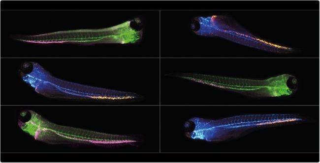
Multiplexed Zebrafish screening in micro plates with Hermes system. Image Credit: IDEA Bio-Medical Ltd.
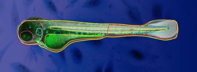
Auto segmentation of Zebrafish organelles empowered by deep learning. Image Credit: IDEA Bio-Medical Ltd.
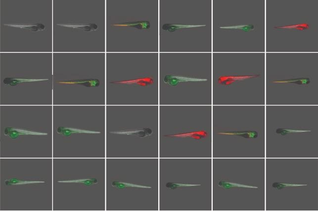
Screen 96 Zebrafish in less than 2 minutes with Hermes imaging system. Image Credit: IDEA Bio-Medical Ltd.