The discovery of fluorescent proteins from many marine organisms has revolutionized scientific research. Scientists use fluorescent proteins in many biological studies, such as those determining cell behaviors in real-time. Also, fluorescent proteins act as a marker that helps study gene expression.
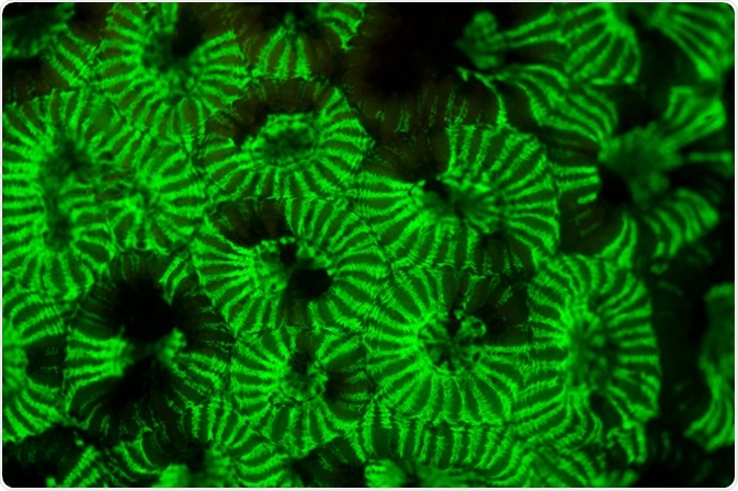
Image Credit: Ethan Daniels/Shutterstock.com
A brief history of fluorescent proteins
Osamu Shimomura, who was a young researcher with an inquisitive mind, tried to understand what was the reason for crystal jelly (Aequorea victoria) glowing bright green when agitated? Shimomura soon isolated the luminescent luciferase enzyme from the jellyfish and realized that in the presence of calcium (present in seawater) it appears as a bright blue light and not green. He hypothesized that jellyfish contain a compound that absorbs blue light and emits green light. He identified this protein and named it Green Fluorescent Protein (GFP).
Later, researchers found that the GFP of the jellyfish contains 238 amino acids. Among these, three amino acids, i.e., numbers 65 to 67, form a structure that emits visible green fluorescent light. GFP present in the jellyfish interacts with another protein, called aequorin, which emits blue light in the presence of calcium. Scientists have also identified the Gfp gene which encodes for GFP.
Upon the discovery of GFP by Shimomura, Martin Chalfie, of Columbia University, tried inserting the DNA sequence that encodes for GFP into the roundworm's DNA. He had successfully expressed GFP along with any gene of interest in the roundworm. The glow served as a marker to identify the location where genes were being expressed. This innovation transformed various invisible processes into visible and opened a new path for biological and medical research.
Advancements in fluorescent proteins
GFP is the most popularly used fluorescent protein that paved a glorious future for exploring cells and their varied mechanisms. GFP can be easily attached to specific proteins and a wide range of enzyme targets. Researchers revealed that cells (express gene products) that are tagged with fluorescent proteins can be visualized with low light intensities for many hours. As this GFP fused product fluoresces, it helps visualize and track known proteins to further understand cellular events.
When a protein is tagged with a specific target, scientists monitor cellular processes within living systems with the help of optical microscopy and other relevant methodology. Recently, scientists have combined various improvements that proved to be invaluable in live-cell imaging experiments, such as the use of ultrafast low light level digital cameras and multi-tracking laser control systems and the GFP with color-shifted genetic derivatives.
One of the limitations of GFP is that it fades too quickly for some research applications. To overcome this drawback, Roger Tsien, a biochemist at UC San Diego, developed new variations of GFP that were brighter and glowed in an array of different colors. He named these variations as “Monomeric banana” and “Tandem dimer tomato”. These variations allowed researchers to monitor multiple cellular processes at the same time with greater precision. They were also used by the researchers at Harvard University to map individual neurons of the mouse brain and create an iconic image called “brainbow”.
Where can you find fluorophore?
Besides Aequorea victoria, scientists have also isolated fluorescent protein from marine sea anemone Discosoma striata and reef corals belonging to the class Anthozoa. These are longer wavelength fluorescent proteins that emit orange and red spectral wavelengths. They have reported that most marine organisms are bioluminescent.
Although many fluorescent proteins are found in sea creatures, far-red and near-infrared molecules tend to come from bacteria. Most luciferase enzymes found in the bioluminescent organisms, except for the bacterial luciferases, require additional luciferin for their emission.
On the contrary fluorescent proteins fluoresce just upon excitation. Some of the advantages of fluorescent protein imaging over luciferase imaging are that it provides brighter signals, substrate independence, availability in multiple colors, and simpler and cheaper equipment requirements.
Recently, several free online resources have become available that help scientists select appropriate fluorescent proteins. Some of the examples of online platforms are FPbase (Lambert), Fluorescence SpectraViewer (Thermo Fisher), and the Fluorescence Spectra Analyzer (BioLegend). These platforms show the excitation and emission curves for hundreds of fluorescent proteins and dyes.
Application of fluorescent proteins
The luciferase enzymes isolated from bioluminescent organisms and fluorescent proteins are used as genetic tracers and biosensors. Researchers at the UC San Diego visualized breast cancer cells in real-time in 2007. As fluorescent proteins can stably infect tumor cells, they are immensely used in cancer research.
Tumor cells can express two or more different colored fluorescent proteins, e.g., the nucleus can be tagged with a green fluorescent protein and the cytoplasm with a red fluorescent protein. This enables imaging of nuclear-cytoplasmic dynamics in a living cell. One of the advantages of this technique is that the tagged tumors and metastases can be visualized non-invasively.
Fluorescent proteins can also be used for in vivo imaging of a single cell. Single-cell imaging can be used to study cancer cell invasion, seeding in distant organs, and dormancy. Also, fluorescent proteins can be used for imaging the efficacy of drug candidates for the treatment of cancer in real-time in mouse models of human cancer.
Photoactivatable fluorescent proteins (PAFPs) is a new tool that helps scientists to visualize the structures and processes in living cells, non-invasively, at the molecular level. PAFPs can follow cancer cells as they seek out blood vessels and spread throughout the body. They are also capable of monitoring how cells manage intracellular debris and preventing premature aging.
Further Reading
Last Updated: Nov 12, 2021