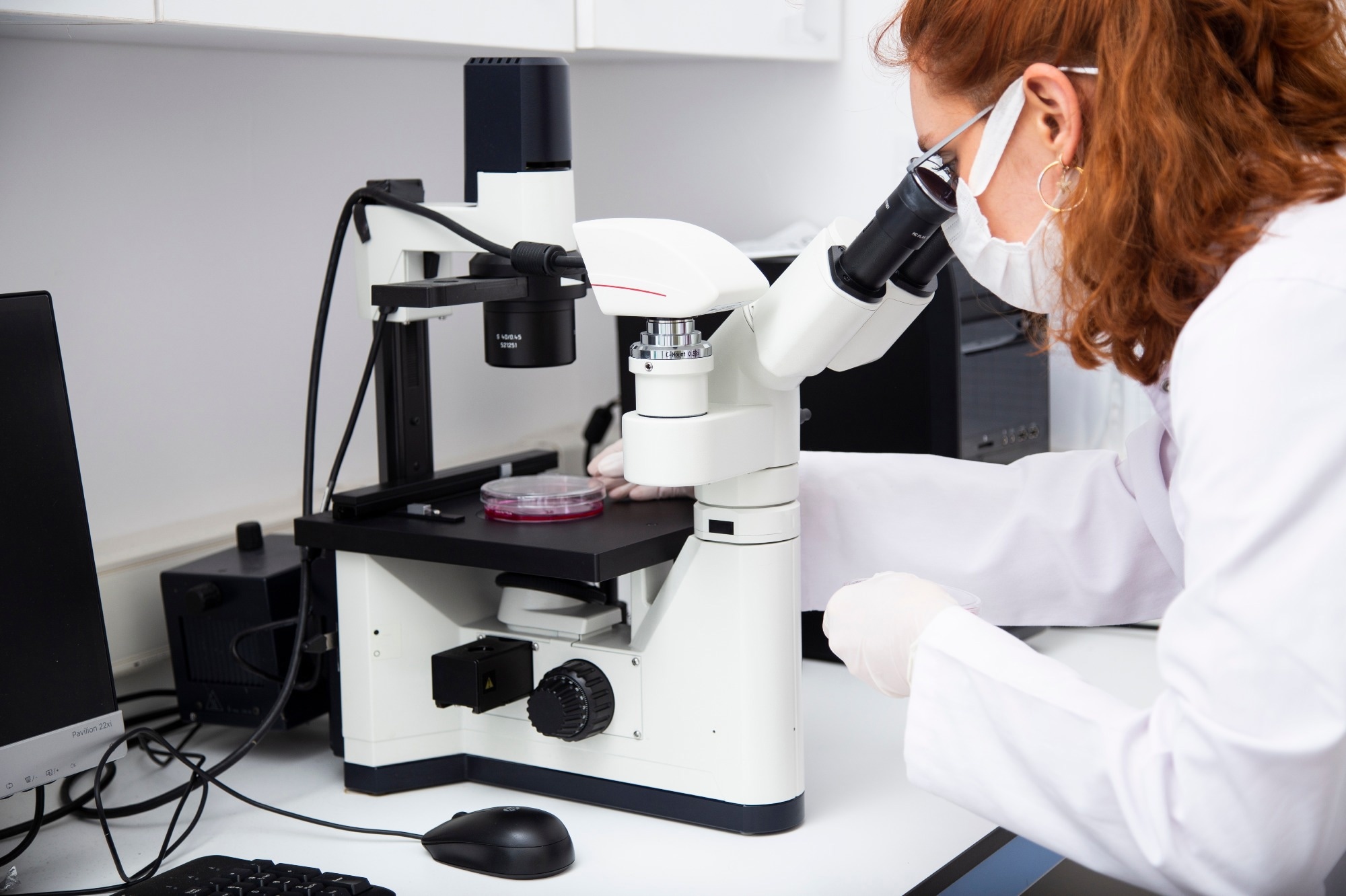
The advent of single-molecule imaging techniques has transformed the understanding of molecular dynamics. These techniques enable the direct observation of individual molecules in real-time, providing unprecedented insights into their behavior and interactions.
Single-molecule imaging is a powerful and versatile tool that allows the study of individual biomolecule dynamics in high detail, providing new insights into molecular processes such as DNA replication, protein-protein interactions, and enzyme kinetics.
 Image Credit: murat photographer/Shutterstock.com
Single-Molecule Imaging: Foundations and Advances
The ability to visualize individual molecules with high spatial and temporal resolution is fundamental to single-molecule imaging. Key techniques include fluorescence microscopy, atomic force microscopy, and super-resolution imaging.
Single-molecule imaging fluorescence microscopy relies on the emission of light from a molecule when excited by a specific wavelength and has been instrumental in providing valuable insights into cellular processes by visualizing the movement of individual molecules within cells.
In particular, single-molecule imaging with total internal reflection fluorescence (TIRF) has become very popular. TIRF microscopy has a high signal-to-noise ratio thanks to its excellent optical sectioning and background suppression. The resulting high sensitivity paves the way for studying complex biological processes such as protein folding and cellular signaling.1
Atomic force microscopy (AFM) creates images based on the forces between a probe and the sample surface. It is suitable for imaging molecules in their native environments with no need for labels or dyes, making it a powerful tool for studying molecular structures and dynamics.2
Super-resolution (SR) fluorescence microscopy overcomes the diffraction limits of conventional optical microscopy and is a promising approach for imaging biological samples. SR imaging techniques such as STORM (stochastic optical reconstruction microscopy) and PALM (photoactivated localization microscopy) have opened up new avenues for studying cellular structures and processes, such as studies of the neuronal synapse.3
Live-Cell Imaging with Super-Resolution Microscopy
Technological Innovations in Single-Molecule Imaging
Advancements in recent years have led to enhanced sensitivity, resolution, and speed. These include the development of novel fluorophores, engineered probes, and imaging platforms, allowing to overcome traditional barriers such as background noise and photobleaching,
Advances in detector technology, labeling methods, and image analysis algorithms have further enhanced the capabilities of single-molecule imaging, contributing to more detailed and accurate observations of molecular dynamics.
Applications of Single-Molecule Imaging in Research
Single-molecule imaging techniques find applications in many disciplines, from biochemistry to genetics and nanotechnology. The ability to visualize protein folding pathways, DNA dynamics, or enzyme reactions provides insights into fundamental biological processes such as DNA replication and protein folding and permits the investigation of complex biomolecular assemblies.
Single-molecule imaging technologies allow direct observation of fluorescently labeled biomolecules in aqueous solutions, enabling the determination of their dynamic interactions and behavior. For instance, single-molecule FRET (smFRET) has been applied to the analysis of the DNA-unwinding mechanisms of various DNA helicases acting on single DNA molecules.4
smFRET can also be used to characterize the transition paths between protein folding and unfolding states. There are reports of the technique being applied to elucidate the conformational dynamics of CRISPR-Cas9, mouse metabotropic glutamate receptor 2 (mGluR2), and the yeast 26S proteasome.1
Researchers used AFM-single-molecule force spectroscopy to analyze chain-folded molecules during single-crystal growth and examine the evolution of the molecular structure over time. A similar approach allows us to characterize the protein folding/unfolding transition state and to clarify the folding process.5
Single-molecule force spectroscopy was used to study the kinetics of the folding steps associated with hormone binding and activation of the glucocorticoid receptor, an important signaling protein, and a prominent drug target.6
Challenges and Solutions in Single-Molecule Imaging
Despite the vast potential of single-molecule imaging and advancements in the field, several limitations persist. One of the major challenges is photobleaching – the irreversible loss of fluorescence caused by repeated excitation – which poses limitations on the observation time.
Another limitation is the need to label biomolecules with fluorophores, which may introduce artifacts or alter the natural behavior of molecules and their functionality. Low signal-to-noise ratios and data complexity also pose challenges that require the integration of experimental techniques with computational modeling and statistical analysis.
Methodological advancements in sample preparation, data acquisition, and image processing have significantly improved single-molecule studies, enabling more accurate interpretations of molecular behavior.
Integrating Single-Molecule Imaging with Other Techniques
Combining high-resolution imaging with complementary analytical techniques enables a more comprehensive understanding of molecular structures and interactions.
For instance, when electron microscopy (which offers extremely high-resolution images of molecular structures) is combined with single-molecule imaging, it is possible to correlate the dynamic information obtained from single-molecule studies with the structural information provided by electron microscopy. This leads to a more complete picture of molecular processes.
Similarly, single-molecule imaging can be combined with mass spectrometry to gain insights into the composition and dynamics of molecular assemblies. Whilst the former technique can reveal the dynamics of a protein complex, the latter can provide information on the identities and quantities of the proteins involved.
Future Directions in Single-Molecule Imaging
Emerging trends in single-molecule imaging include the development of novel fluorescent probes and the integration of machine learning for data interpretation, with the potential to further revolutionize the understanding of molecular mechanisms.
Future investigations should aim to enhance labeling techniques (including genetically encoded tags and chemical modification approaches) with focus on achieving higher efficiency, specificity, and minimal disruption to the molecule functionality.
Simultaneous imaging of multiple species or different structural components within complex systems is another area of interest that is due to expand. Future endeavors will involve designing and synthesizing fluorophores with narrower emission spectra to minimize spectral overlap, and refining spectral separation techniques.
Conclusion
Single-molecule imaging and the integration with other analytical techniques have emerged as crucial tools to shed light on the dynamics of individual molecules and gain a more comprehensive understanding of molecular processes.
As further research leads to the development of improved and new techniques, single-molecule imaging will be able to drive discoveries and innovations with implications for medicine, biotechnology, and various other scientific fields.
References and Further Reading
- Colson, L., Kwon, Y., Nam, S., Bhandari, A., Maya, N. M., Lu, Y. & Cho, Y. (2023). Trends in Single-Molecule Total Internal Reflection Fluorescence Imaging and Their Biological Applications with Lab-on-a-Chip Technology. Sensors (Basel), 23.10.3390/s23187691.
- Li, S., Wang, X., Li, Z., Huang, Z., Lin, S., Hu, J. & Tu, Y. (2021). Research progress of single molecule force spectroscopy technology based on atomic force microscopy in polymer materials: Structure, design strategy and probe modification. Nano Select, 2, 909-931.https://doi.org/10.1002/nano.202000235. Available: https://onlinelibrary.wiley.com/doi/abs/10.1002/nano.202000235
- Gagliano, G., Nelson, T., Saliba, N., Vargas-Hernández, S. & Gustavsson, A. K. (2021). Light Sheet Illumination for 3D Single-Molecule Super-Resolution Imaging of Neuronal Synapses. Front Synaptic Neurosci, 13, 761530.10.3389/fnsyn.2021.761530.
- Takahashi, S., Oshige, M. & Katsura, S. (2021). DNA Manipulation and Single-Molecule Imaging. Molecules, 26.10.3390/molecules26041050.
- Lv, C., Gao, X., Li, W., Xue, B., Qin, M., Burtnick, L. D., Zhou, H., Cao, Y., Robinson, R. C. & Wang, W. (2014). Single-molecule force spectroscopy reveals force-enhanced binding of calcium ions by gelsolin. Nature Communications, 5, 4623.10.1038/ncomms5623. Available: https://doi.org/10.1038/ncomms5623
- Suren, T., Rutz, D., Mößmer, P., Merkel, U., Buchner, J. & Rief, M. (2018). Single-molecule force spectroscopy reveals folding steps associated with hormone binding and activation of the glucocorticoid receptor. Proc Natl Acad Sci U S A, 115, 11688-11693.10.1073/pnas.1807618115.
Last Updated: May 15, 2024