Western blotting is a laboratory technique used to qualify or quantify specific protein content within a sample. It is a well-established technique that uses antibodies to discern the presence of certain proteins within an electrophoresed sample that does not need to be purified beforehand, thus making it quicker and more cost-effective than other protein identification techniques.
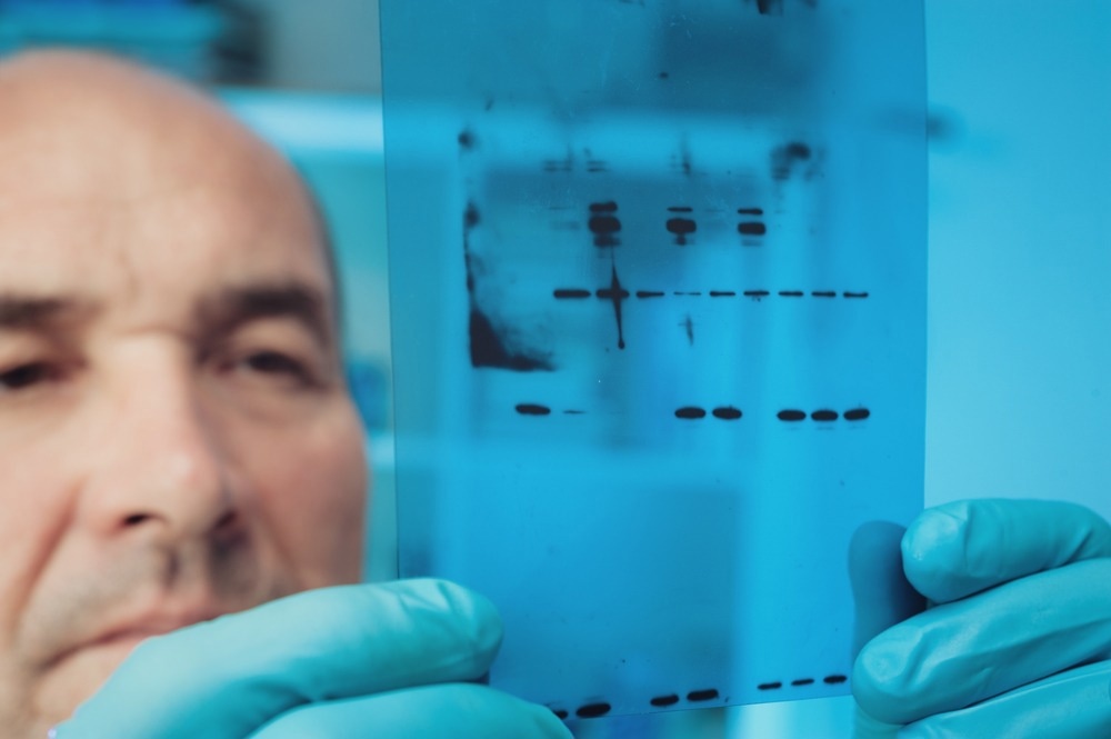
Image Credit: tilialucida/Shutterstock.com
Western blotting is a technique credited to the laboratory of Harry Towbin in 1979 and is named based on a play on Southern blotting, a similar technique used to detect DNA that Edwin Southern invented. Though individual techniques may vary, the overall original procedure remains the same – sample electrophoresis, transfer of proteins from a gel to a membrane, detection of those proteins using primary antibodies, and visualization of proteins using secondary antibodies. Technique specifics can be optimized based on the manufacturer's recommended protocols and trial and improvement within the lab setting.
Preparing Western Blot Membranes
Firstly, samples are lysed to release proteins and centrifuged to allow the separation of proteins from other elements of whole-cell samples, i.e., DNA and mitochondria. Purified proteins can also be used, for example, in a pull-down experiment, if desired. Protein samples are then prepared for electrophoresis by dilution to desired levels depending on the protocol being used and adding a sample dye so electrophoretic movement can be visualized.
If using a Western blot for quantification, it is crucial that all protein samples are prepared to the same content concentration, which requires a step beforehand of sample quantification using an assay compared to a standard curve, such as a bicinchoninic acid (BCA) assay (for cell quantification) or bovine serum albumin (BSA) assay (for purified protein quantification).
Once the samples are prepared, they can be loaded into an electrophoresis gel, typically a sodium dodecyl sulphate–polyacrylamide gel electrophoresis (SDS-PAGE) gel, alongside a molecular weight ladder. This gel is then run in an electrophoresis chamber filled with a running buffer for a set time at a current or voltage that can be protocol-specific. This can be varied based on the molecular weight of the target proteins, the density of the gel, and several other factors.
Once the gel has run to a satisfying level, the next step is to transfer the proteins from the gel to a membrane (commonly nitrocellulose), which is most frequently achieved by wet transfer electroblotting. The gel and membrane are prepared in a 'sandwich' consisting of sheets of filter paper on either side of the gel and membrane, encased in two sponges and held together in a plastic casing. The prepared 'sandwich' must also be rolled or pressed to remove bubbles, as these will cause interference in membrane visualization, and be kept wet with transfer buffer to prevent damage to gels that can become brittle if allowed to dry. However, dry transfer techniques can be used, typically in conjunction with an automated blotting system. The transfer then occurs in an electrophoresis chamber, with the gel orientated to allow the negatively charged proteins to shift towards the positive anode, transferring from the gel to the membrane, again in a process that may differ depending on circumstances.
Once transferred, it is important that membranes are blocked using a protein solution containing, for example, milk or BSA to prevent non-specific antibody binding.
Protein Visualisation
Primary antibodies must then be incubated with the prepared membrane to visualize protein content within the sample. The primary antibody used must be specific to the target protein, i.e., an anti-ATPase antibody if ATPases are the target proteins of the study. The animal-of-origin of these antibodies must also be known to allow secondary antibody binding in the subsequent steps of the process.
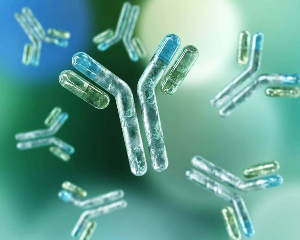
Image Credit: ustas7777777/Shutterstock.com
Immunofluorescence
If immunofluorescence detection is used, antibodies from a separate animal source against a loading control protein should also be incubated in addition to the target protein. Common loading controls or housekeeping proteins used are actin and tubulin, as they are universal to most human cell samples. However, other loading control techniques are in development, such as using total protein staining to better quantify protein content. These loading controls are important, especially for quantification blots, as they allow the comparison of quantities of the target protein to other proteins, so changes under specific experimental conditions can be evaluated.
Following primary antibody incubation and an optional additional blocking process to prevent background interference, secondary antibody incubation can occur. Two separate secondary antibodies are used for the target protein and loading control protein, as they must bind to the primary antibody's animal-specific, or Fc, region. For example, if a rabbit antibody was used for the target protein and a mouse for the loading control, then anti-rabbit and anti-mouse secondary antibodies must be used accordingly. The secondary antibodies used allow the visualization of samples due to the presence of a wavelength-specific fluorescent molecule. The fluorescence wavelength of the target and loading control antibodies must be different to allow them to be discerned.
Protein presence can then be visualized using an immunofluorescence detection machine, of which there are several available. The blots can then be seen and analyzed using appropriate software.
Non-immunofluorescence
Not all Western blots are visualized using secondary immunofluorescent antibodies due primarily to high costs. If using an alternative visualization technique, membranes can only be blotted using one primary antibody at a time, and a secondary antibody conjugation to another visualization protein, often horseradish peroxidase, which can be detected using an enhanced chemiluminescence reagent. This means that no loading control will be seen, so membranes can be stripped and re-blotted to see a loading control after visualization, or multiple gels of the exact same loading conditions run concurrently to allow separate blots with separate antibodies.
If a membrane is going to be stripped, it is recommended that the proteins are fixed to it by dipping in methanol before blotting, as this reduces the risk of proteins being lost during a stripping procedure.
Sources:
- Moritz CP (2017) Tubulin or Not Tubulin: Heading Toward Total Protein Staining as Loading Control in Western Blots. Proteomics 17(20). https://doi.org/10.1002/pmic.201600189
- Oh K (2021) Technical Considerations for Contemporary Western Blot Techniques. Methods Mol Biol 2261:457–479. https://doi.org/10.1007/978-1-0716-1186-9_29
- Pillai-Kastoori L, Schutz-Geschwender AR, Harford JA (2020) A systematic approach to quantitative Western blot analysis. Anal Biochem 593:113608. https://doi.org/10.1016/j.ab.2020.113608
- Towbin H, Staehelin T, Gordon J (1979) Electrophoretic transfer of proteins from polyacrylamide gels to nitrocellulose sheets: procedure and some applications. Proc Natl Acad Sci U S A 76(9):4350–4354. https://doi.org/10.1073/pnas.76.9.4350
Further Reading
Last Updated: Jan 25, 2023