Using cutting-edge video microscopy, researchers from the Walter and Eliza Hall Institute (WEHI) have successfully observed the molecular intricacies of how malaria parasites enter red blood cells—a critical phase in the disease.

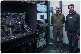
Dr. Geoghegan (left) and Dr. Whitehead (right) with the lattice light-sheet microscope. Image Credit: Walter and Eliza Hall Institute.
The researchers captured high-resolution films of individual parasites entering the red blood cells with the help of a customized lattice light-sheet microscope—the first in Australia—and observed the cellular and molecular alterations that take place throughout this process. The study has yielded crucial new data on malaria parasite biology that could be useful for developing the much-needed new antimalarial drugs.
Ms. Cindy Evelyn, Dr. Niall Geoghegan, Dr. Lachlan Whitehead, Professor Alan Cowman, and Dr. Kelly Rogers conducted the study, which was recently published in the Nature Communications journal.
At a glance
- A sophisticated microscope technology, known as lattice light-sheet microscopy, was utilized to capture complete, real-time footage of the malaria parasite attacking the red blood cells.
- The study has uncovered important steps in the parasite invasion process, which is a pivotal stage in the malaria life cycle and underpins several malaria symptoms.
Focusing on a deadly parasite
Malaria is known to be a mosquito-borne disease that claims the lives of around 400,000 people worldwide each year. Many of the devastating symptoms of malaria are caused by the invasion and development of the Plasmodium parasite in the red blood cells of an infected person, stated Dr. Rogers, the director of WEHI's Centre for Dynamic Imaging.
Understanding in better detail exactly how the parasite invades red blood cells may reveal new ways to stop this stage of the parasite life cycle, potentially leading to much-needed new therapies.”
Dr Kelly Rogers, Head, Centre for Dynamic Imaging, Walter and Eliza Hall Institute
“We used microscopy—specifically a state-of-the-art approach, lattice light-sheet microscopy (LLSM)—to follow the intricate cellular and molecular changes that occur when the malaria parasite invades red blood cells. We captured the first-ever high-resolution, real-time, and dynamic views of the parasite in action,” Dr. Rogers added.
According to Ms. Evelyn, who began the study as an Honors student, the study revealed many formerly unknown aspects of parasite invasion.
The videos we recorded showed the 'push and pull' interactions as the parasite landed on the red blood cell, and then entered the cell in an enclosed chamber – called a vacuole – where it grew and multiplied. There has long been contention in the field about whether the vacuole is derived from the parasite or the host cell. Our research resolved this question, revealing it was created from the red blood cells membrane.”
Ms Cindy Evelyn, Centre for Dynamic Imaging, Walter and Eliza Hall Institute
A majority of antimalarial vaccines and therapies target the initial binding of malaria to red blood cells.
By visualizing these processes in more detail, our research may contribute in several ways to the development of new antimalarial therapies. For example, now that we know that the parasite vacuole relies on components of the red blood cell membrane, it might be possible to target these components with medicines to disrupt the parasite life cycle. This host-directed approach could be one way to bypass the malaria parasite’s propensity to rapidly develop drug resistance.”
Dr Kelly Rogers, Head, Centre for Dynamic Imaging, Walter and Eliza Hall Institute
“LLSM may also have applications for observing the specific steps of parasite invasion that are blocked by potential new drugs—an area we are now very interested in pursuing,” Dr. Rogers added.
New views of cells
LLSM is a cutting-edge imaging technology that allows scientists to observe organs and cells in incredible detail and in real-time. According to Dr. Geoghegan, LLSM has altered the way cells could be examined.
“In the past, the choice of microscope for an experiment had to be a compromise between capturing a lower resolution video, which revealed dynamic processes like shape changes or movement, and capturing much higher-definition still images, which provided much more detail about how the cells and molecules are functioning,” added Dr. Geoghegan.
“LLSM allows us to obtain high-resolution videos of cells, which has been a game-changer for many fields of biological research. We custom built a LLSM at WEHI – the first version of this technology in Australia. This groundbreaking microscopy has enabled us to progress in multiple areas of research, including this malaria study. To achieve this, we brought together a multidisciplinary team with expertise in physics, engineering, and biology – and the results of this current study have vindicated our approach,” concluded Dr. Geoghegan.
Microscopic CCTV reveals secrets of malaria invasion
Video Credit: Walter and Eliza Hall Institute.
Source:
Journal reference:
Geoghegan, N. D., et al. (2021) 4D analysis of malaria parasite invasion offers insights into erythrocyte membrane remodeling and parasitophorous vacuole formation. Nature Communications. doi.org/10.1038/s41467-021-23626-7.