 By Pooja Toshniwal PahariaReviewed by Lexie CornerMay 12 2025
In a recent study published in the Journal of Clinical and Translational Hepatology, researchers explored the importance of the intestinal transmembrane protein TM6SF2 in the progression of metabolic dysfunction-associated steatotic liver disease (MASLD), particularly when triggered by a high-fat diet (HFD).
Researchers highlight a growing body of evidence linking gut health to liver function, pointing to TM6SF2 as a potential therapeutic target in MASLD management.

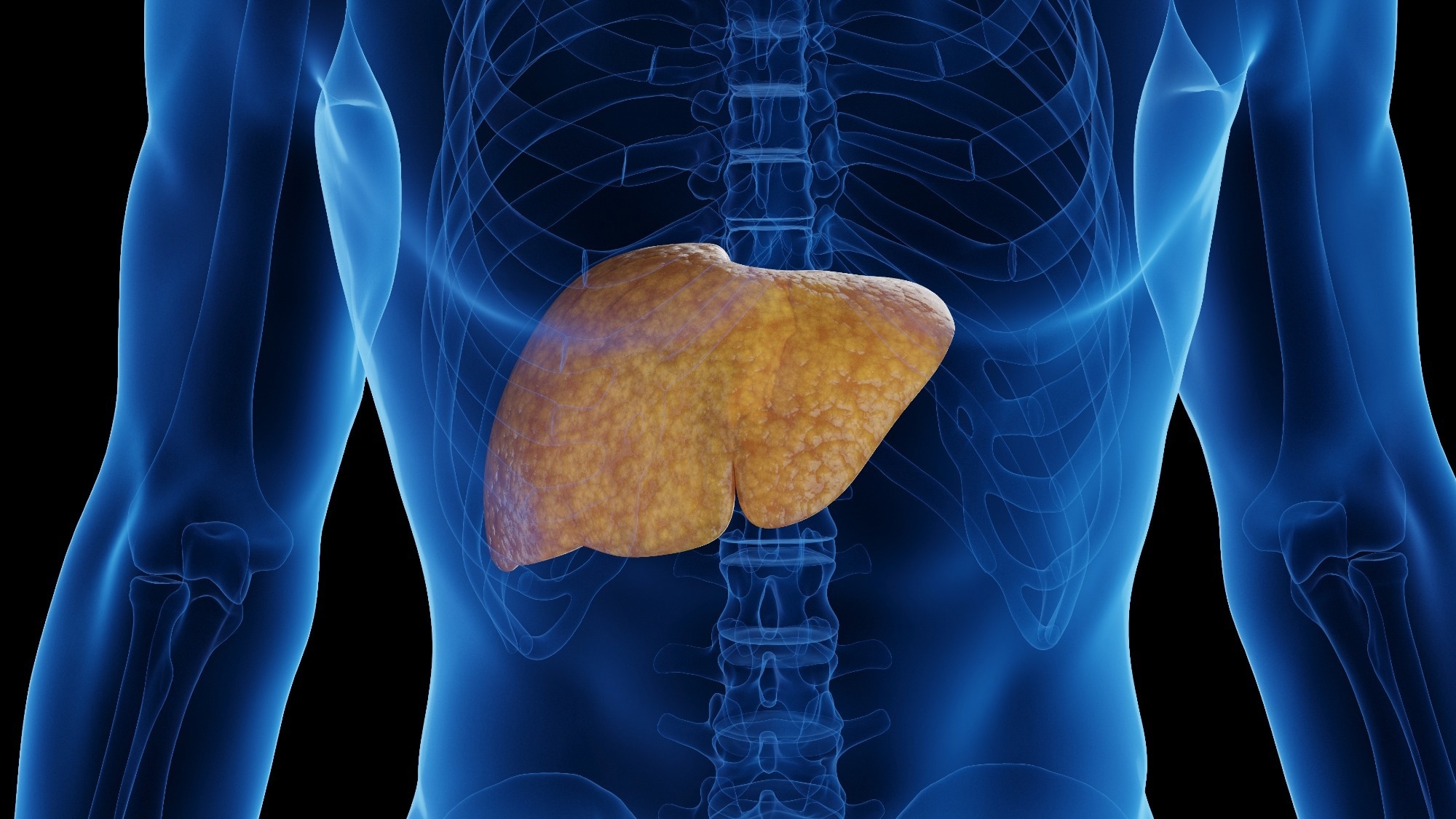
Image Credit: 3dMediSphere/Shutterstock.com
Previously known as non-alcoholic fatty liver disease (NAFLD), MASLD is the most prevalent form of chronic liver disease. Its development is influenced by multiple factors, including inflammation, endoplasmic reticulum (ER) stress, oxidative damage, and lipotoxicity.
More recently, studies have also shown that gut microbial imbalances—known as dysbiosis—can worsen MASLD by altering microbial composition and metabolism in ways that intensify liver pathology.
TM6SF2, a transmembrane protein expressed primarily in the gut and liver, has been identified as a contributor to hepatic triglyceride buildup, especially when the E167K mutation is present. This mutation has been associated with an increased risk of MASLD.
About the Study
To understand the role of TM6SF2 in MASLD, researchers created a gut-specific TM6SF2 knockout mouse model (TM6SF2 GKO) and fed them a high-fat diet. These mice were compared to wild-type (CON) mice also fed an HFD, as well as to those on a standard control diet (CD), over a 16-week period.
The high-fat diet consisted of 60 % fat, 20 % protein, 20 % carbohydrates, and 5.21 kcal/g, whereas the control diet contained 10 % fat, 20 % protein, 70 % carbohydrates, and 3.85 kcal/g.
Using CRISPR/Cas9 gene-editing technology, the team knocked out TM6SF2 in the gut. They extracted RNA from tissue samples to synthesize complementary DNA and amplify TM6SF2 expression, confirmed through RNA sequencing, Western blot, and real-time PCR. To profile the gut microbiome, DNA was extracted from fecal samples for 16S rRNA sequencing, followed by partial least squares discriminant analysis (PLS-DA) to assess inter-group microbial differences.
Serum samples were analyzed for markers of liver function and lipid metabolism, including AST, ALT, triglycerides (TG), low-density lipoprotein (LDL), high-density lipoprotein (HDL), and total cholesterol (TC). Metabolite profiling was conducted using ultra-high-performance liquid chromatography coupled with mass spectrometry (UHPLC-QE-MS).
The researchers also examined lipid accumulation in the liver and intestines. Oil Red O staining was used to visualize lipid droplets, while hematoxylin and eosin (H&E) staining revealed liver steatosis.
Key Findings
Mice on the high-fat diet exhibited significant lipid accumulation in both the liver and intestines. This effect was amplified in the TM6SF2 GKO-HFD group, which showed notably higher lipid levels than their wild-type counterparts.
Elevated serum levels of AST, ALT, HDL, TG, and TC in these mice further confirmed worsened liver function. Visual inspection of the liver revealed enlargement, discoloration, and the presence of lipid droplets—all signs of advanced steatosis.
The gut microbiome also changed dramatically in the TM6SF2 GKO-HFD group. These mice had a lower Firmicutes/Bacteroidetes (F/B) ratio, reduced species richness, and lower alpha diversity compared to controls. Taxonomic analysis found higher levels of Clostridia_UCG_014, Candidatus Arthromitus, and Desulfovibrio, alongside reduced populations of Ruminococcustorques and Intestinimonas.
PLS-DA further supported the distinct microbial profiles across groups. Tissue staining with Alcian Blue showed a reduction in goblet cells in the intestines of HFD-fed mice, implying increased intestinal permeability. This could allow bacterial metabolites to reach the liver through the portal vein, potentially triggering inflammation, fibrosis, and liver damage.
Serum metabolomics identified significant shifts in compound levels in TM6SF2 GKO-HFD mice. Seventeen metabolites, including lysophosphatidylcholine 18:0 (LPC 18:0), were elevated, while 22 others, such as phenylsulfate, were reduced.
These results suggest that TM6SF2 affects lipid metabolic pathways under HFD conditions. Several of these metabolites, including LPC 18:0 and oleoylcarnitine, emerged as potential biomarkers for MASLD severity.
Pathway analysis via the Kyoto Encyclopedia of Genes and Genomes (KEGG) showed downregulation of the sphingolipid pathway, along with upregulation of pathways linked to choline metabolism and efferocytosis, further implicating LPC 18:0 in disease progression.
Download your PDF copy now!
Conclusion
This study underscores the importance of TM6SF2 in maintaining gut-liver metabolic balance. When TM6SF2 is depleted in the gut, high-fat diets can more readily disrupt lipid metabolism, gut microbial diversity, and liver function, ultimately increasing the risk of MASLD. These insights could help identify new biomarkers and therapeutic strategies to better diagnose and manage the disease.
Journal Reference
Li-Zhen Chen, Yu-Rong Wang, Zhen-Zhen Zhao, Shou-Lin Zhao, Cong-Cong Min, and Yong-Ning Xin. Intestinal Depletion of TM6SF2 Exacerbates High-fat Diet-induced Metabolic Dysfunction-associated Steatotic Liver Disease through the Gut-liver Axis. J Clin Transl Hepatol, 2025. DOI: 10.14218/JCTH.2024. 004072025, https://www.xiahepublishing.com/2310-8819/JCTH-2024-00407