Microgels are colloidal particles consisting of cross-linked, 3D polymer networks. These particles can expand or contract considerably when affected by environmental stimuli, including pH levels, ionic strength, temperature, and light.
This ability to expand and contract enables the triggered release of encapsulated bioactive molecules. In addition, these particles defend the loaded molecules against degradation, and they can also enhance their physical stability.
Consequently, microgels have been identified as good candidates for tuneable drug delivery systems. Moreover, as they can be functionalized in a relatively straightforward way, with potential applications, including tissue engineering and biosensors, microgels could be a promising prospect when considering material development applications.1,2
Measurements and results
Borro et al.3 utilized Small Angle X-ray Scattering (SAXS) on the BioXolver L to acquire structural information on microgels that consisted of biodegradable polymer alginate, packed with polymyxin B – a model antimicrobial peptide.
They carefully examined how preparation techniques and the concentration of the ionic crosslinker Ca2+ and polymyxin B provoked the microgel structure. The SAXS data sets Borro et al. gathered were analyzed with a core-shell model fit, whose parameters are illustrated in Figure 1. This generated information on the mesh size (ξ) of the polymer network and the dimension of the dense core (Rc ), as well as data on the microgel’s (2σ) fuzzy outer layer. For a detailed analysis of the study, see the literature by Borro et al.3
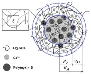
Figure 1. Core-shell model used for fitting of the microgel SAXS data. ξ: mesh size of the polymer network, Rc: radius of the dense core, σ: the thickness of the shell, Rg: radius of gyration. Image Credit: Xenocs
It was discovered that the mesh size ξ (Figure 2, A) and the size of the fuzzy layer 2σ (Figure 2, B) were for the most part unaffected by the amount of polymyxin B. Comparatively, growth of the microgel core Rc (Figure 2, B) was witnessed when more peptide was loaded, which correlated with the notion that polymyxin B is located within the particle’s core.
With the introduction of increased levels of crosslinker, a smaller mesh size presented with a denser polymer network (Figure 2, C). An overall reduction in the size of the particle was observed (Figure 2, D). SAXS data exhibited that the core and the fuzzy layer both diminished in size with increased levels of crosslinking.
 and crosslinker (CaCl2) respectively.")
Figure 2. Structural parameters of the microgels, extracted from the SAXS data as a function of the amount of peptide-loaded (Polymyxin) and crosslinker (CaCl2) respectively. Image Credit: Xenocs
Summary
The robotic loading of the BioXolver facilitates automated investigations into a greater number of samples. Therefore, it is an advantageous system optimized for the study of microgels, which generally necessitates the investigation of a wide variety of conditions.
References
- Agrawal, G. & Agrawal, R. Functional Microgels: Recent Advances in Their Biomedical Applications. Small 2018, 14, e1801724.
- Plamper, F. & Richtering, W. Functional Microgels and Microgel Systems. Accounts Chem. Res. 2017, 50, 131–140.
- Borro, B. et al. Microfluidics-Based Self-Assembly of Peptide-Loaded Microgels: Effect of Three Dimensional (3D) Printed Micromixer Design. J. Colloid Interf. Sci. 2019, 538, 559-568.
About Xenocs
Xenocs is a supplier of x-ray scattering equipment (SAXS/WAXS) for characterizing the nanostructure and morphology of materials at the nanoscale. Such equipment is used for the research, development, and production of advanced materials in various domains such as nanoparticles & colloïds, polymer, food science, cosmetics, or biostructural research.
Created in 2000 as a spin-off from Institute Laue Langevin, the company started activity offering its customers innovative X-ray sources, optics, and collimation solutions for X-ray characterization of nanomaterials and nanostructures.
Xenocs delivered its first x-ray scattering equipment in 2008, setting new standards for the possibilities of using SAXS in the laboratory for characterization at the nanoscale.
Working closely with both academic and corporate customers, Xenocs has continuously focused on providing value through performance and ease of use, while also creating a global sales and service organization.
In 2016, Xenocs bought Saxslab with operations in Denmark and in Massachusetts, making it the leading provider of SAXS equipment worldwide. Xenocs headquarters are located in France, and the company has subsidiaries in the USA, Denmark, and Singapore as well as a strong network of local contacts.
Sponsored Content Policy: AZO Life Science publishes articles and related content that may be derived from sources where we have existing commercial relationships, provided such content adds value to the core editorial ethos of AZO Life Science, which is to educate and inform site visitors interested in medical research, science, medical devices, and treatments.