Electron microscopy has become a vital tool in structural biology, enabling researchers to visualize biological macromolecules at near-atomic resolution. Recent advances have transformed it from a low-resolution imaging method into a powerful technique for elucidating molecular mechanisms, driving progress in drug discovery, disease research, and more.
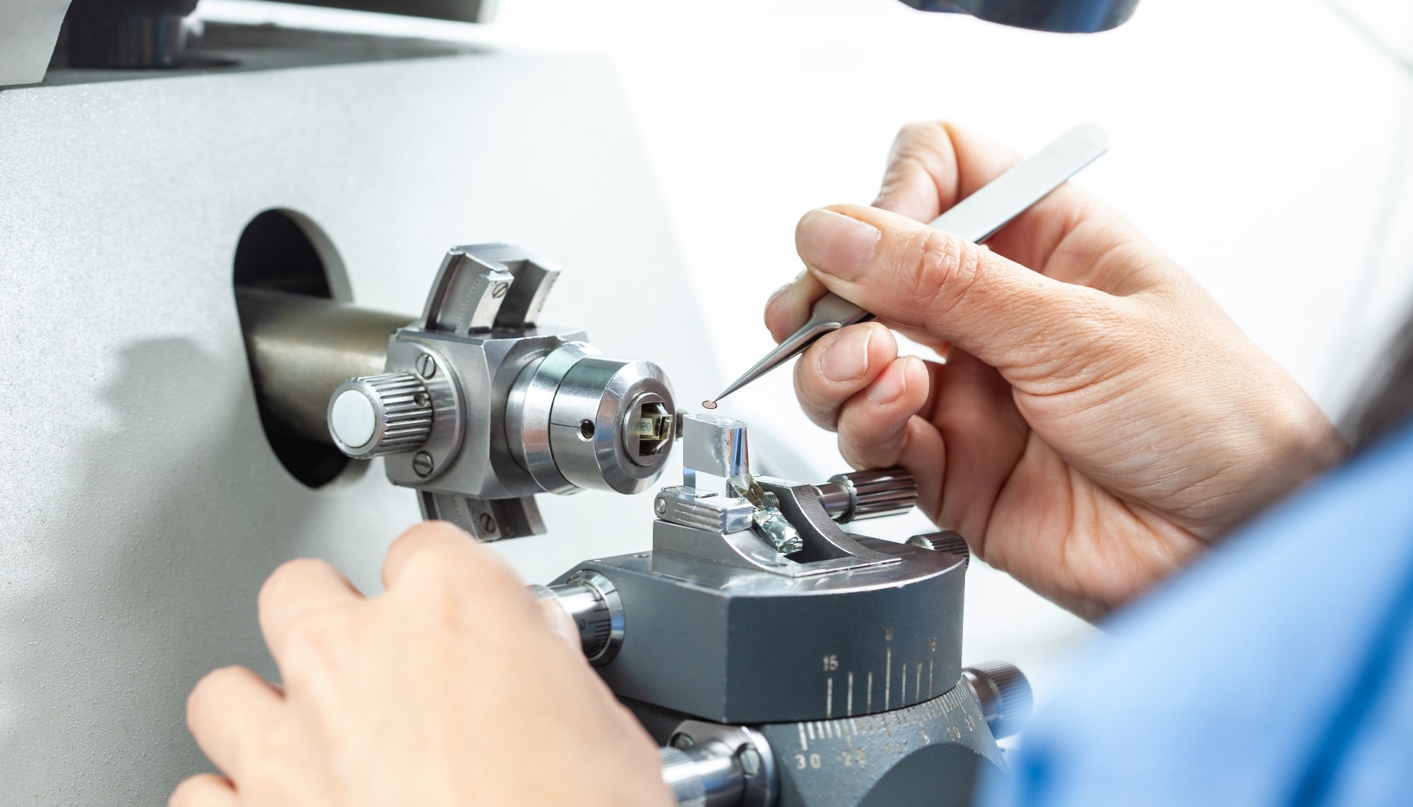 Image Credit: Anamaria Mejia/Shutterstock.com
A Brief History of Electron Microscopy
By 1900, optical microscopy had reached its theoretical limit, with visible light microscopes achieving magnifications of up to 1000x and resolving details up to approximately 0.2 micrometres (2000 angstroms). Looking to take resolution further, Carl Zeiss introduced the first commercial ultraviolet microscope in 1904, which doubled the resolution attainable with visible light. However, this new microscope was still limited by the wavelength of light.
In 1931, Ernst Ruska and Max Knoll changed the game. Microscopy was revolutionized by the introduction of electrons, exploiting their shorter wavelength to achieve far greater resolution. This breakthrough led to the development of the first transmission electron microscope (TEM), which generated images by transmitting electrons through ultrathin samples, uncovering structural details far beyond the reach of optical microscopy.
A decade later, Ruska designed the scanning electron microscope (SEM), using a focused beam of electrons to scan the surface systematically. The resulting image was generated by detecting scattered electrons, providing exceptional depth and clarity in information on surface morphology and composition.
The next major advancement came in 1986 when the digital microscope was introduced in Japan, which allowed real-time image capture and computer-based analysis. Modern systems now integrate high-definition digital displays, allowing direct visualization without the need for external computing equipment.1
From 2D Imaging to 3D Resolution with Cryo-Electron Microscopy
One of the most significant limitations in microscopy was its inability to accurately convert low-resolution two-dimensional images into high-fidelity three-dimensional structures. Unable to do this restricted the study of biological macromolecules in their native conformations.
Jacques Dubochet, Joachim Frank, and Richard Henderson overcame this in the 80s with their novel cryogenic vitrification technique that involved rapidly cooling biological samples to approximately −180°C in liquid ethane. This process transformed water into an amorphous, glass-like state rather than crystalline ice, preserving the specimen’s hydrated and near-native architecture while minimizing radiation damage during imaging. Their method, known as cryo-electron microscopy (cryo-EM), enabled the visualization of biomolecules with exceptional structural fidelity, transforming high-resolution biological imaging.
Progress continued through the 90s and 00s, with the most significant advances jumping off from 2013, when electron detectors were introduced into cryo-electron microscopy (cryo-EM). They replaced conventional charge-coupled device (CCD) cameras, providing enhanced sensitivity, superior image quality, and beam-induced motion correction.2
Since then, single-particle cryo-EM has become a crucial technique for elucidating the structures of large macromolecular complexes that are challenging to crystallize, such as ribosomes, membrane proteins, and viruses. It requires no crystal formation, uses minimal sample quantities, and can capture multiple conformational states within a single dataset.
The most recent developments have achieved sub-1.5 Å resolution, meaning even individual atoms can be visualized, and highly accurate models of protein architectures can be created. This has led to landmark studies, including the structural characterization of the ribosome, spliceosome, and nuclear pore complex, each contributing essential insights into the structural foundations of cellular function.2
Cryo-Electron Tomography: Visualizing Molecular Architecture In Situ
Cryo-electron tomography (cryo-ET) is an extension of cryo-electron microscopy (cryo-EM) that enables the three-dimensional visualization of non-repetitive structures within their native cellular environments.
Unlike single-particle analysis, which averages data from thousands of identical molecules, cryo-ET captures a tilt series of images from a single specimen at multiple angles. Computational algorithms reconstruct these projections into a three-dimensional tomogram that reveals the specimen’s volumetric architecture.
The resulting 3D architectures are particularly valuable for examining pleomorphic structures, cellular organization, and macromolecular assemblies in situ. They enable the visualization of structures such as organelles, cytoskeletal frameworks, and membrane systems without the distortions caused by chemical fixation or staining. This has provided critical insights into biological processes such as viral entry, bacterial division, and synaptic organization.
Recent developments in cryo-focused ion beam (cryo-FIB) milling have expanded its applicability to thicker samples, including entire cells and tissues, by thinning vitrified specimens into electron-transparent lamellae of 100 to 300 nm while preserving their structural context. When combined with sub-tomogram averaging, cryo-ET can attain nanometer-scale resolution, effectively bridging the gap between single-particle cryo-EM and cellular imaging.3
The potential of cryo-electron tomography
Video Credit: Max Planck Institute of Molecular Physiology
Correlative Light and Electron Microscopy: Bridging Scales
A drawback of conventional electron microscopy is that it is unable to specifically identify molecular components or dynamic biological events within the dense, complicated expanse of cellular ultrastructures.
While electron microscopy can offer excellent spatial resolution, it lacks the molecular specificity provided by fluorescence-based analysis, making it challenging to pinpoint proteins, organelles, or transient cellular processes within a sample.
Correlative light and electron microscopy (CLEM) was developed to overcome this limitation by integrating fluorescence microscopy with electron microscopy. In this multimodal approach, fluorescence imaging is first employed to label and visualize specific molecules or regions of interest using fluorescent probes, genetically encoded tags, or immunolabeling techniques.
Once identified, the same specimen is imaged with an electron microscope, and computational tools correlate the fluorescence signals with ultrastructural features, preserving spatial accuracy between both modalities.4,5
Biological Impact of Electron Microscopy Advances
Advancements in electron microscopy have transformed structural and cellular biology by enabling atomic-level visualization of complex biomolecular assemblies.
Cryo-electron microscopy (cryo-EM) has become a central technique in investigating membrane proteins, which represent approximately 30 % of the human proteome and constitute the majority of therapeutic drug targets. It has enabled high-resolution structural analysis of G-protein coupled receptors (GPCRs), ion channels, and transporters, revealing mechanisms of molecular recognition and signaling that have advanced structure-based drug design.
It has also elucidated the organization of essential cellular machines such as the 26S proteasome, the spliceosome, and the nuclear pore complex, providing detailed insight into protein degradation, RNA processing, and nucleocytoplasmic transport.6,7
The COVID-19 pandemic highlighted the impact of cryo-EM in structural virology, when structures of the SARS-CoV-2 spike protein were determined within weeks of genome sequencing, facilitating the rapid design of vaccines and antibody development.8
Further results have been achieved in neuroscience, where cryo-ET and CLEM have revealed the intricate organization of synaptic components, including neurotransmitter receptors, scaffolding proteins, and vesicular networks, uncovering principles of synaptic transmission and plasticity. These methods have also resolved the molecular architecture of axonal transport systems, dendritic spines, and myelin sheaths, establishing direct correlations between molecular structure and neuronal function.9
Challenges and Limitations
Despite major advancements, there are several technical and methodological constraints that continue to limit electron microscopy.
The quality of vitrified specimens remains a critical factor in determining resolution, yet achieving optimal ice thickness, particle distribution, and orientation remains difficult, particularly for membrane proteins that tend to adopt preferred orientations.
Radiation damage also limits achievable resolution, as even under cryogenic conditions, electron exposure must be carefully controlled to maintain structural integrity. This issue is especially pronounced in cryo-ET, where multiple image acquisitions reduce the allowable dose per projection.
Computational analysis presents further challenges due to the high data volume and the complexity of image-processing algorithms, which require expert validation to prevent artifacts. Structural heterogeneity within biological samples adds further difficulty, requiring advanced classification methods to distinguish different molecular states.
Moreover, size remains a limiting factor: particles smaller than about 40 kDa yield insufficient contrast for accurate alignment, while larger specimens may exceed the thickness or field-of-view limits of TEM.10,11
Download your PDF now!
Future Developments and Prospects
Electron microscopy is progressing toward greater resolution, speed, and accessibility through technological and computational innovations.
Future direct electron detectors are expected to enhance sensitivity and temporal precision, providing faster data collection and improved visualization of dynamic biological processes. Advancements in phase plate technology could increase image contrast of small particles, expanding the range of biomolecular structures that can be resolved at ultra-high resolution.
More and more, artificial intelligence and machine learning are transforming data processing pipelines by automating quality assessment, particle selection, and classification. Computing approaches help to improve image reconstruction through noise suppression and better handling of structural heterogeneity, allowing reliable analysis from smaller or lower-quality datasets.
Time-resolved electron microscopy has emerged in recent times to overcome the static limitations of conventional methods, using microfluidic vitrification and ultrafast pulsed electron beams to capture transient molecular states and dynamic structural changes on picosecond to millisecond timescales.12,13
Conclusion
Electron microscopy has advanced significantly over the past decade, transitioning from a niche imaging approach to a central method for resolving macromolecular structures at atomic precision. As innovations continue to make the technology faster, more accessible, and more powerful, its role in biomedical research will only continue to grow, enabling the translation of molecular-level understanding into targeted therapeutic and diagnostic advancements.
References and Further Reading
- Shaber, L. (2018). The History of the Electron Microscope. https://www.thermofisher.com/blog/materials/the-history-of-the-electron-microscope/
- Chui, G. (2022). Cryogenic electron microscopy (cryo-EM): amazing views of life’s machinery. https://www6.slac.stanford.edu/research/slac-science-explained/cryo-em
- Tocheva, E. I., Li, Z., & Jensen, G. J. (2010). Electron Cryotomography. Cold Spring Harbor Perspectives in Biology, 2(6), a003442. https://doi.org/10.1101/cshperspect.a003442
- Yang, J., Vrbovská, V., Franke, T., Sibert, B., Larson, M., Hall, A., Rigort, A., Mitchels, J., & Wright, E. R. (2023). Integrated Fluorescence Microscopy (iFLM) for Cryo-FIB-milling and In-situ Cryo-ET. BioRxiv, 2023.07.11.548578. https://doi.org/10.1101/2023.07.11.548578
- Cognigni, F., Miraglia, L., Contessi, S., Biancardi, F., & Rossi, M. (2023). Correlative Light and Electron Microscopy (CLEM): A Multifaceted Tool for the Study of Geological Specimens. Journal of Experimental and Theoretical Analyses, 1(2), 74-85. https://doi.org/10.3390/jeta1020006
- Robertson, M. J., Meyerowitz, J. G., & Skiniotis, G. (2021). Drug discovery in the era of cryoEM. Trends in Biochemical Sciences, 47(2), 124. https://doi.org/10.1016/j.tibs.2021.06.008
- Wehmer, M., & Sakata, E. (2016). Recent advances in the structural biology of the 26S proteasome. The International Journal of Biochemistry & Cell Biology, 79, 437-442. https://doi.org/10.1016/j.biocel.2016.08.008
- Cortese, M., & Laketa, V. (2021). Advanced microscopy technologies enable rapid response to SARS‐CoV‐2 pandemic. Cellular Microbiology, 23(7), e13319. https://doi.org/10.1111/cmi.13319
- Nature Conferences. (2025). Advances in electron microscopy drive diverse scientific fields. https://doi.org/10.1038/d42473-024-00385-9
- Cianfrocco, M. A., & Kellogg, E. H. (2020). What Could Go Wrong? A Practical Guide To Single-Particle Cryo-EM: From Biochemistry To Atomic Models. Journal of Chemical Information and Modeling, 60(5), 2458. https://doi.org/10.1021/acs.jcim.9b01178
- Ling, W. L., Kimura, Y., Han, Y., & Li, Y. (2023). Editorial: Recent advances and challenges in electron microscopy characterizations of radiation-sensitive nanoparticles. Frontiers in Chemistry, 11, 1171240. https://doi.org/10.3389/fchem.2023.1171240
- Berkeley, R. F., Cook, B. D., & Herzik, M. A. (2024). Machine learning approaches to cryoEM density modification differentially affect biomacromolecule and ligand density quality. Frontiers in Molecular Biosciences, 11, 1404885. https://doi.org/10.3389/fmolb.2024.1404885
- Alcorn, F. M., Jain, P. K., & M., R. (2023). Time-resolved transmission electron microscopy for nanoscale chemical dynamics. Nature Reviews Chemistry, 7(4), 256-272. https://doi.org/10.1038/s41570-023-00469-y
Last Updated: Oct 9, 2025