The ImageXpress® Pico Automated Cell Imaging System is more than just a digital microscope; it combines high-resolution imaging with advanced analysis. The automated imager offers a detailed portfolio of preconfigured protocols for cell-based assays to lessen the learning curve and allow users to start running experiments rapidly, whether users are running fluorescence imaging or brightfield assays.
With features such as Digital Confocal* 2D on-the-fly deconvolution, Autofocus, Live Preview, multi-wavelength cell scoring, and optional IN Carta® Image Analysis Software workflow, the ImageXpress Pico helps users advance their discoveries in a small, affordable imager.
Get Started Quickly
The entire lab can optimize its digital microscopy with the icon-driven, user-friendly CellReporterXpress® Image Acquisition and Analysis Software. Users can start capturing and analyzing images with little to no training.
Do More Than Cell Counting
Over 25 preconfigured templates optimized for several cell-based experiments, including mitochondrial evaluation, apoptosis, 3D cell models, live cell/timelapse, multiwavelength cell scoring, and neurite tracing, are available to help users expand their assays.
Automate Imaging Affordably
There is no need to travel to the core lab to have the samples run. The system’s low cost enables scientists to have the convenience of automated imaging and analysis on their lab bench. The system can be customized to fit the research needs with options such as Digital Confocal, environmental control, and z-stack acquisition.
ImageXpress Pico Automated Imaging System Virtual Tour. Image Credit: Molecular Devices UK Ltd
Features
Multiple Imaging Modes
The ImageXpress Pico system has objectives ranging in magnification from 4× to 63× and can perform colorimetric, brightfield, fluorescence, or Digital Confocal 2D on-the-fly deconvolution imaging.
Preconfigured Analysis Protocols
Over 25 preconfigured analysis protocols are available, ranging from simple cell counting to advanced neurite tracing analysis. Analysis parameters can be optimized using features such as the click-to-find tool by simply clicking on a few cells that suit specific criteria.
Plate-to-Individual Cell View
Data can be viewed at various levels, ranging from a plate overview to individual cells. A wide range of data visualization tools, from the plate to the cellular level, enable users to gain as much information as possible from their images and assays.
Z-stack acquisition
Using z-stack acquisition, users can produce sharper images for more accurate segmentation. To acquire more detail than a single slice, take a series of images at different focal points. Users can include all slices in the final projection or choose which slices to include.
Quickly and Easily Identify Regions of Interest
The Live Preview mode makes it easier to identify regions of interest, allowing users to pan around the sample and modify the focus interactively with a virtual joystick, saving both time and effort.
Environmental control
The onboard environmental system, which includes humidity, CO2, and O2 control, can be used to run multi-day, time-lapse, and live cell assays. The software, which has been optimized to prevent z-drift, also provides real-time monitoring of the environmental state, optimizing assay conditions.
Experience the Powerful Combination of the ImageXpress Pico and CellReporterXpress
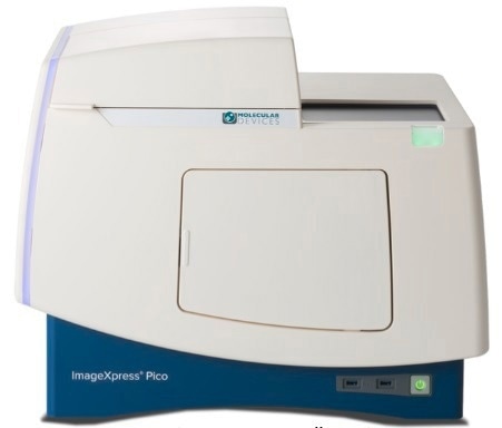
ImageXpress Pico Automated Cell Imaging System. Image Credit: Molecular Devices UK Ltd
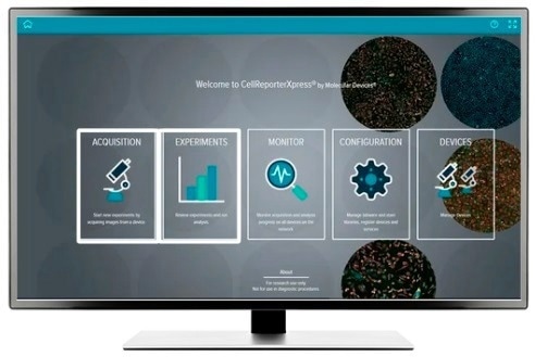
CellReporterXpress Image Acquisition and Analysis Software. Image Credit: Molecular Devices UK Ltd
CellReporterXpress Software for ImageXpress Pico
Easy-to-Learn Software Optimized for Automated Digital Microscopy
A straightforward, user-friendly interface for conducting quantitative analysis on images obtained from automated microscopy, including Digital Confocal 2D real-time deconvolution, Autofocus, and Live Preview for a sophisticated region of interest identification.
The software allows for distributed image analysis for higher throughput and is ideal for scaling digital microscopy imaging with slides or microplates. An icon-driven, linear workflow provides a streamlined user experience with a variety of predefined protocols.
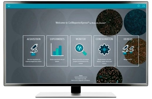
Image Credit: Molecular Devices UK Ltd
IN Carta® Image Analysis Software
Go from Assay to Insights Quickly and Reliably
Image analysis and phenotypic profiling workflows are simplified by powerful analytics coupled with an intuitive user interface. Advanced features enable users to analyze data in 2D, 3D, and 4D at scale and deliver real-time insights without needing complex pre- or post-processing operations.
The precision of the image analysis workflows with the SINAP deep-learning module is greatly enhanced, and users can see that segmentation is not a problem. Machine learning can be used to conduct a complex phenotypic analysis within a user-friendly Phenoglyphs module.
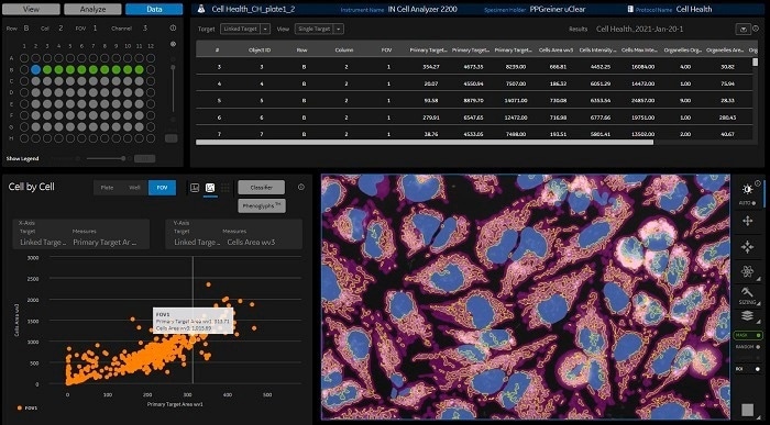
Image Credit: Molecular Devices UK Ltd
Tailored Laboratory Automation Solutions With Robotics, Incubators, and Software
The research environment is changing rapidly, and today’s researchers require improved laboratory automation and simplified remote access. By combining the ImageXpress® Pico Automated Cell Imaging System with the S-LABTM plate handler from PAA and the SCILA incubator from Inheco, users can achieve increased productivity, lower costs, and consistent performance.
Automated ImageXpress Pico workflow in action. Image Credit: Molecular Devices UK Ltd