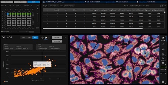
IN Carta® Image Analysis Software helps to solve complex image analysis issues using cutting-edge Artificial Intelligence (AI) to transform images into outcomes that can be easily interpreted.
User-friendly workflows mean results can be obtained quicker from 2D, 3D, and 4D experiments. Using the Custom Module Editor application, users can characterize highly tailored image analysis protocols enabling them to achieve robust outcomes — even for complicated assays — then swiftly review, visualize, and interact with the analysis outcomes. IN Carta software does the heavy lifting so users can concentrate on their research.
Powerful
Guided workflows and scalable batch processing help to increase productivity and reduce the time to answer. Experiments can be set up quickly, and analysis of multiple wells is run in parallel.
Insightful
Machine learning allows users to leverage more information and increase accuracy when analyzing high-content screening data to confidently enable new discoveries.
Intuitive
Modern user experience and cutting-edge technology help minimize the software learning curve, removing the barriers to productivity.
IN Carta Image Analysis Software. Image Credit: Molecular Devices UK Ltd.
Features
Deep Learning
Using the SINAP module, users can enhance the specificity of the image analysis workflows. It is based on deep learning-based image analysis, which results in robust segmentation for virtually all biological structures.
Focused Worklists
Browse through a parent directory and populate a worklist with image datasets of interest or use search to locate a dataset of interest.
AI-Powered Data Analytics
Users are able to leverage the power of machine learning without having to be trained data scientists.
The user-friendly workflow helps users identify and quantify phenotypic changes. Users can also explore data and reveal insights from complex datasets and identify novel and unexpected phenotypes at the click of a few buttons.
Customization
Browse and analyze images from experiments, produce image analysis protocols of varied complexities, and include on-demand data classification. With 360 ̊ data linking among data tables, images, and charts, users are able to analyze their results.
3D analysis
The 3D application Custom Module Editor offers unparalleled flexibility in segmenting complicated biological structures. Image datasets can be attained in 3D/4D (timelapse 3D), and customized image analysis routines can be made within a guided workflow.
Batch analysis and monitoring
Using one or more analysis protocols, explore multiple experiments in batch analysis mode. Track the status of any submitted task and supervise their development in real-time.
IN Carta SINAP
SINAP is a module that uses deep learning algorithms to improve the accuracy and reliability of high-content screening assays during segmentation — the first step in the analysis pipeline. It offers improved object detection compared to conventional image analysis methods.
Deep learning models can be easily tailored within a user-friendly tool, so that any novel biological objects can be segmented efficiently. The quantitative information that is extracted from segmented objects is more precise, so errors are not propagated down the analysis pipeline.
With SINAP, Segmentation Is Not a Problem
- Accurate – Deep learning can maintain accuracy across difficult to segment samples including confluent cells, low signal-to-noise samples and transmitted light images.
- Reliable – SINAP models can account for high phenotypic variability.
- Flexible – A single workflow can handle a selection of imaging modalities and applications.
- Accessible – The trained model learns to segment from scientist’s drawing on the image instead of asking a deep-learning expert to develop a new model and regulate multiple parameters
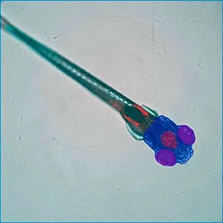
Customized SINAP deep-learning models segment specific regions (whole body, head, eyes, brain) of a zebrafish embryo in transmitted light image. Courtesy of Guo Lab, UCSF. Image Credit: Molecular Devices UK Ltd.
IN Carta Phenoglyphs
The IN Carta® Phenoglyphs™ Software Module employs a special combination of supervised and unsupervised machine learning to measure phenotypical modifications. With several hundreds of cellular characteristics that can be examined simultaneously, an all-inclusive phenotypic profile is made and can be applied across a complete screening workflow.
This multivariate way of classification offers a precise definition of object populations that allows users to resolve subtle phenotypic alterations made by genetic modification or drug treatment. It can be used across several biological targets alongside cells, organoids, spheroids, etc.
- Comprehensive – A data-driven approach that starts with unsupervised clustering to find patterns in the data and highlight subpopulations without prior knowledge of what phenotypes may exist.
- Optimized workflow – Classification is achieved by just confirming or correcting the algorithm's predictions until it learns the appropriate behavior.
- Robust – The committed machine learning algorithm determines the ideal set of descriptive characteristics to eliminate overfitting of the subsequent classification model.
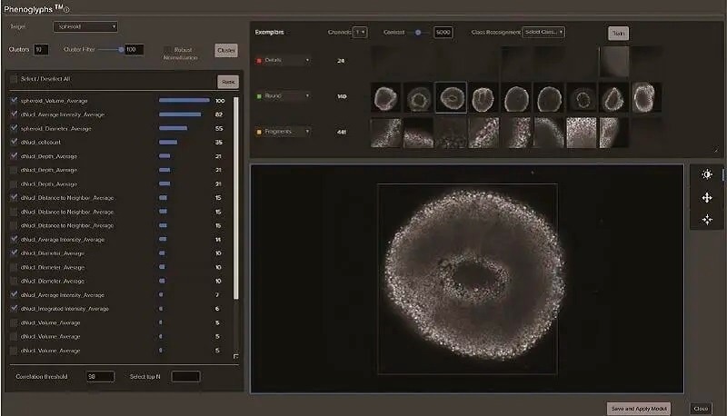
Classification of spheroids formed from HCT116 cells. Spheroids were stained with Hoechst 33342 to visualize nuclei. 3D stacks were collected over time. Image Credit: Molecular Devices UK Ltd.
IN Carta CustomModule Editor 2D and 3D
- Develop easy step-by-step custom analysis
- Identify objects localized within defined biological compartments
- Customize object classification and segmentation
- Only report the measurements needed for an assay of interest
- Examine live imaging data
- Strong reconstruction of 2D segmentation into volume for 3D assays (3D application only)
- True 3D segmentation (3D application only)
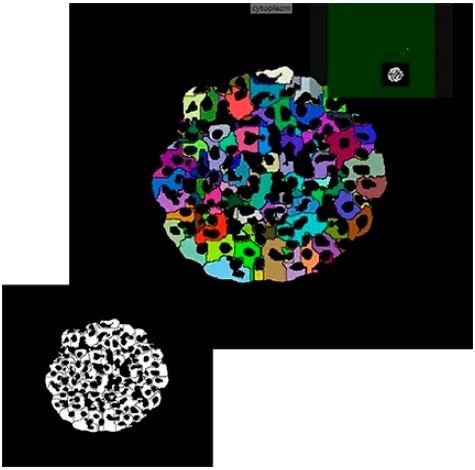
Example segmentation of iPSC-derived hepatocyte spheroids in Custom Module Editor. Image Credit: Molecular Devices UK Ltd.
ImageXpress® Confocal HT.ai High-Content Imaging System
Powerful Multi-Laser Light Sources, a Deep Tissue Penetrating Confocal Disk Module, Water Immersion Objectives, and Modern Machine Learning Analysis Software.
- Best-suited for highly complicated 3D and cell-based assays.
- Seven-channel high-intensity lasers produce brighter images with a higher signal-to-background ratio.
- Spinning confocal disk technology for deeper tissue penetration produces sharper images with increased resolution.
- Water immersion objectives provide up to quadruple the signal at lower exposure times for higher image clarity and sensitivity.
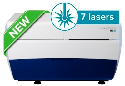
Image Credit: Molecular Devices UK Ltd.
StratoMineR Advanced Cloud-Based Analytics
Generate Clear, Deep Data From Complex Datasets
Efficient and intuitive workflows enable users to port high-content imaging data straight into StratoMineR, where it is employed to produce rich, interactive visualizations with advanced data mining approaches. If employed with IN Carta Image Analysis Software, it offers robust, quantitative results from complicated biological images and datasets using cutting-edge AI technology. Employ all high-content data to define, discover, and examine Phenotypes.
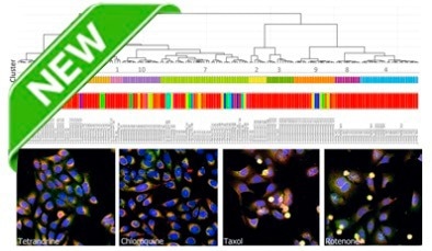
Image Credit: Molecular Devices UK Ltd.