AZoLifeSciences talks to Dr. Ian Wong about a new imaging technique that maps forces that cell clusters exert on their surroundings.
Why is it important to map the 3D forces exerted by tiny cell clusters?
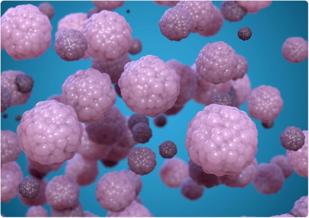
Multicellular clusters mimic the structure and functional behavior of larger tissues, which can deform the surrounding extracellular matrix during wound healing and tumor invasion.
What is Traction Force Microscopy (TFM)?
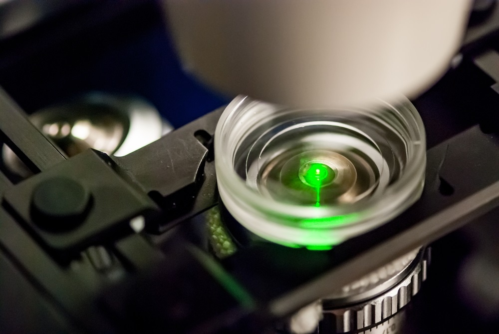
Traction force microscopy is an imaging technique to visualize how soft materials are deformed, based on the motion of embedded tracer particles.
 Image Credits: Micha Weber / Shutterstock.com
Please describe the new technique you have developed.
We embedded individual cells into a soft biomaterial comprised of two types of proteins – collagen and silk, which is decorated with fluorescent tracer particles.
As the cells adhered and migrated, they deform this biomaterial, which results in measurable changes in the positions of these tracer particles.
How did your team go about developing this technique?
This was a collaboration led by my very talented Ph.D. student, Susan Leggett, who had exceptional skills at cell biology and confocal microscopy, working with another Ph.D. student Mohak Patel, who worked on mechanical engineering and computer vision.
They went back and forth to perform some exquisitely sensitive measurements of how cell-generated forces, which they then analyzed.
What were the challenges in determining behaviors within cell clusters?
One challenge is that these clusters are very heterogeneous, so these behaviors can vary considerably from one cluster to the next. It took a lot of careful exploration of the data for us to find meaningful patterns from one experiment to the next.

Image Credit: Christian Zuppinger/Shutterstock.com
What is Displacement Arrays of Rendered Tractions (DART) and how was this developed?
We observed that multicellular clusters deform three-dimensional biomaterials using forces that vary considerably in different regions, which does not occur for individual cells.
We developed a technique to profile each cluster based on how it pulls, pushes, and twists in different regions in its surrounding volume.
How did streamlining the image processing provide benefits for imaging multiple clusters?
Typically, TFM experiments only analyze one cell at a time, partly because the experiments are optimized for certain microscope configurations, and partly because these particle motions are time-consuming to analyze.
We now show that we can analyze a large number of clusters in parallel, which allows us to greatly accelerate how experiments are performed.
How could this method be applied to improve understanding of tissue formation, wound healing and tumor spreading?
Since these clusters mimic larger tissues, we have tested how clusters respond to different biochemical treatments or drugs that enhance or inhibit cell migration. These could predict how these treatments would work to accelerate wound healing or impede tumor invasion.
How could the technique be used to study organoids and what benefits could this bring?
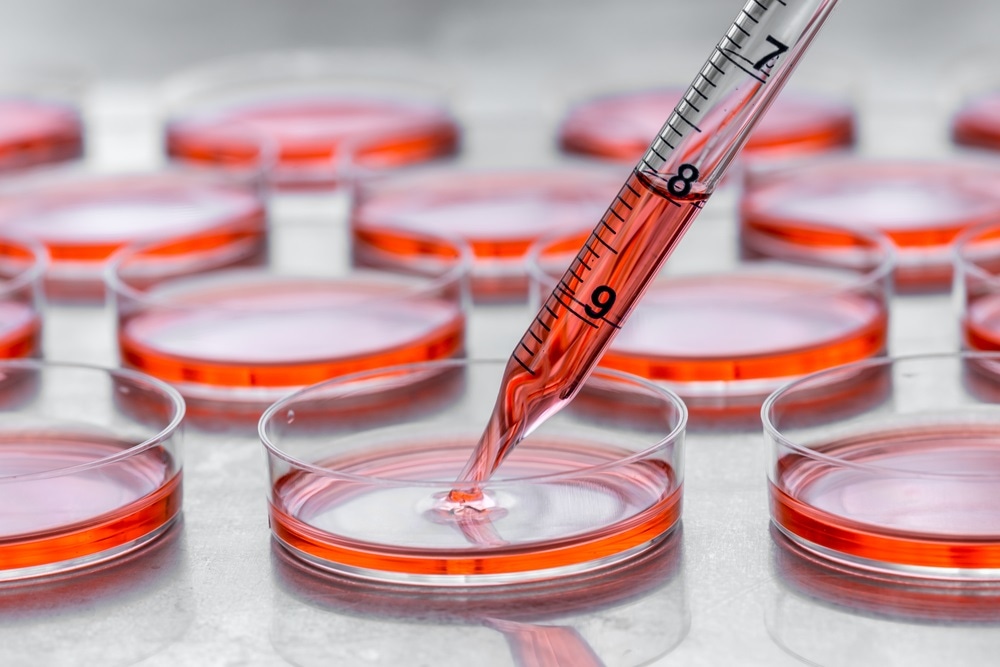
Organoids are based on primary stem cells that can organize into organ-like architecture when cultured outside the body. Organoids have generated much interest as a way to experimentally predict how human patient cells respond to drug treatment.

Image Credits: Hakat / Shutterstock.com
Your team has made the code behind this technique freely available. What do you hope other researchers will be able to utilize your technique for?
We anticipate that this technique can be utilized to investigate how human cells migrate in a variety of contexts, including immune cells, stem cells, or cells that organize into blood vessels.
What are the next steps in your research?
We are particularly interested in extending these techniques to human patient-derived cancer cells, but also in increasing the complexity of the biomaterial to better mimic the extracellular matrix in the body.
Where can readers find more information?
Beri, P. et al. (2018). Biomaterials to model and measure epithelial cancers. Nature Reviews Materials. DOI: https://doi.org/10.1038/s41578-018-0051-6
Polacheck, W.S. and Chen, C.S. (2017). Measuring cell-generated forces: a guide to the available tools. Nature Methods. DOI: https://doi.org/10.1038/nmeth.3834
Leggett, S.E. et al (2020). Mechanophenotyping of 3D multicellular clusters using displacement arrays of rendered tractions. Proceedings of the National Academy of Sciences, USA. DOI: 10.1073/pnas.1918296117
About Dr. Ian Wong

Ian Y. Wong is an Assistant Professor of Engineering and of Medical Science at Brown University.
He received a Ph.D. in Materials Science and Engineering from Stanford University and completed postdoctoral training in bioengineering at Harvard Medical School from 2010-2013. He has been recognized with an NSF Graduate Research Fellowship, a Damon Runyon Cancer Research Fellowship, and the Brown University CareerLAB Pierrepont Prize for Advising.