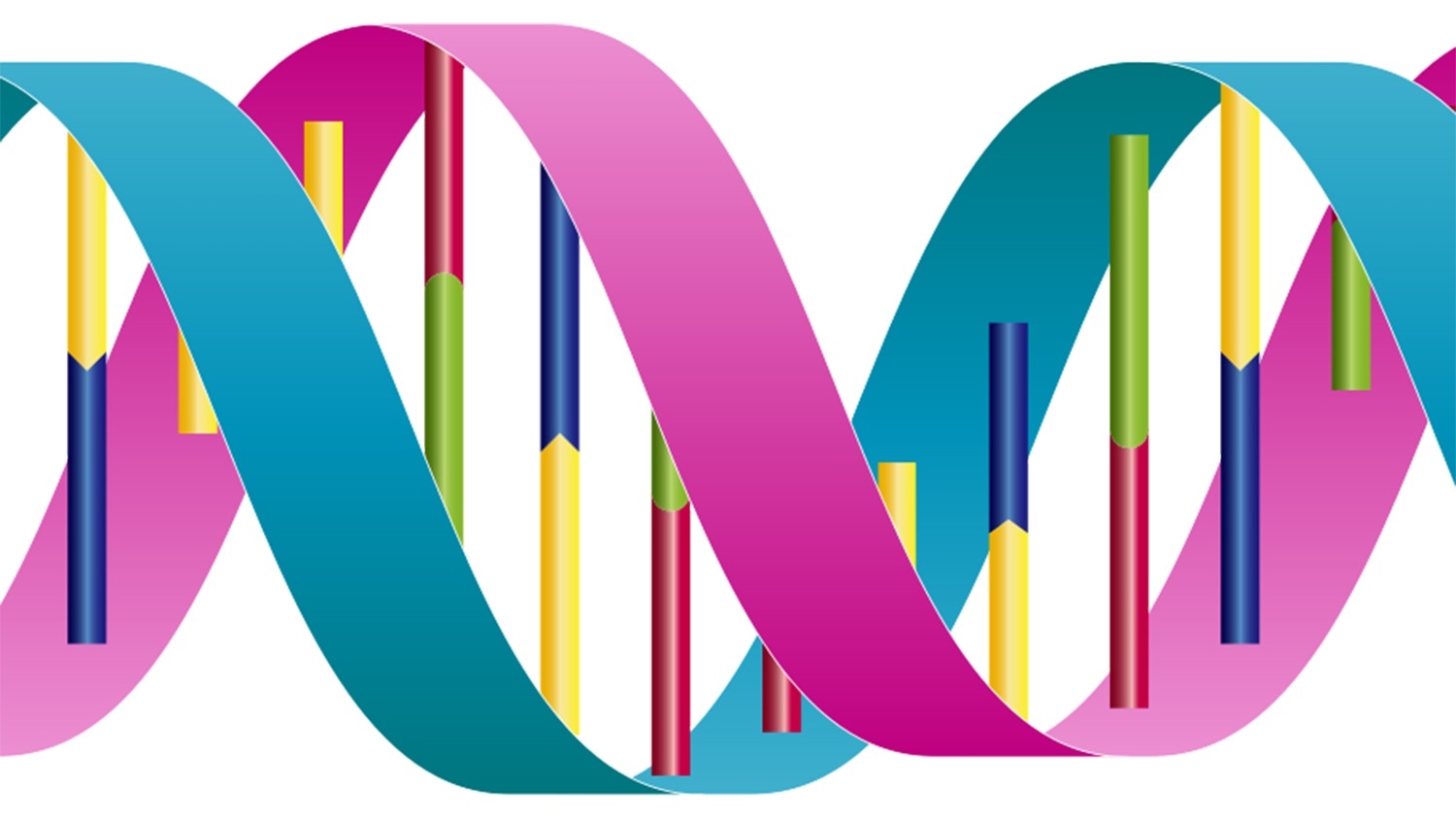
Image Credit: DataBase Center for Life Science. Shared under a Creative Commons license.
Whenever a cell is ready to divide, the DNA also divides, with the double helix “unzipping” into two distinct backbones. New nucleotides—thymine, adenine, guanine, or cytosine—are packed into the gaps on the other part of the backbone, combining with their counterparts (cytosine with guanine and adenine with thymine) and replicating the DNA to create a copy for the new as well as old cells.
A majority of the time, the nucleotides are a perfect match but at times—about 1 time in 10 million—there is inconsistency.
Although mismatches are rare, the human genome contains approximately six billion nucleotides in every cell, resulting in approximately 600 errors per cell, and the human body consists of more than 37 trillion cells. Consequently, if these errors go unchecked they can result in a vast array of mutations, which in turn can result in a variety of cancers, collectively known as Lynch Syndrome.”
Dorothy Erie, Study Corresponding Author and Chemistry Professor, Department of Chemistry, University of North Carolina at Chapel Hill
Erie is also a member of the Lineberger Comprehensive Cancer Center in the University of North Carolina.
To initiate repair of these mismatches, the two proteins, MutL and MutS, work together. The MutS protein slides along the recently produced side of the DNA strand after it is replicated and proofreads it. Upon finding a mismatch, the MutS protein locks into place on the error site and recruits the MutL protein to come and join it.
The MutL protein marks the newly developed strand of DNA as defective and signals a different kind of protein to ingest the part of the DNA that contains the error. Subsequently, the nucleotide matching restarts, filling the gap again.
The whole process decreases the replication errors by about a 1000 times, acting as one of the best defenses of the body against genetic mutations that may result in cancer.
We know that MutS and MutL find, bind, and recruit repair proteins to DNA. But one question remained—do MutS and MutL move from the mismatch during the repair recruiting process, or stay where they are?”
Keith Weninger, Study Co-Corresponding Author and Biophysicist, North Carolina State University
Weninger is also the university faculty scholar at North Carolina State University.
In two distinct papers that were published in the Proceedings of the National Academy of Sciences, Weninger and Erie investigated both bacterial and human DNA to obtain a deeper structural and temporal picture of what actually occurs when the MutL and MutS proteins engage in mismatch repair.
With the help of non-fluorescent and fluorescent imaging techniques, including optical spectroscopy, atomic force microscopy, and tethered particle motion, the scientists identified that the MutL protein “freezes” the MutS in place at the mismatch site, creating a stable complex that remains in that vicinity before repair can occur.
The complex seems to reel in the DNA around the mismatch too, protecting and marking the DNA area until repair can take place.
Due to the mobility of these proteins, current thinking envisioned MutS and MutL sliding freely along the mismatched strand, rather than stopping. This work demonstrates that the process is different than previously thought.”
Keith Weninger, Study Co-Corresponding Author and Biophysicist, North Carolina State University
“Additionally, the complex’s interaction with the strand effectively stops any other processes until repair takes place. So the defective DNA strand cannot be repacked into a chromosome and then carried forward through cell division,” Weninger concluded.
Source:
Journal reference:
Hao, P., et al. (2020) Recurrent mismatch binding by MutS mobile clamps on DNA localizes repair complexes nearby. Proceedings of the National Academy of Sciences. doi.org/10.1073/pnas.1918517117.