In a recent study published in Frontiers in Oral Health, researchers developed advanced oral biofilm models to replicate bacterial dysbiosis associated with peri-implantitis in vitro. The findings revealed that dysbiotic shifts were primarily driven by bacterial species selection, with cultivation conditions playing a secondary but still noticeable role. They occurred without external stimuli, closely mimicking natural oral environments.
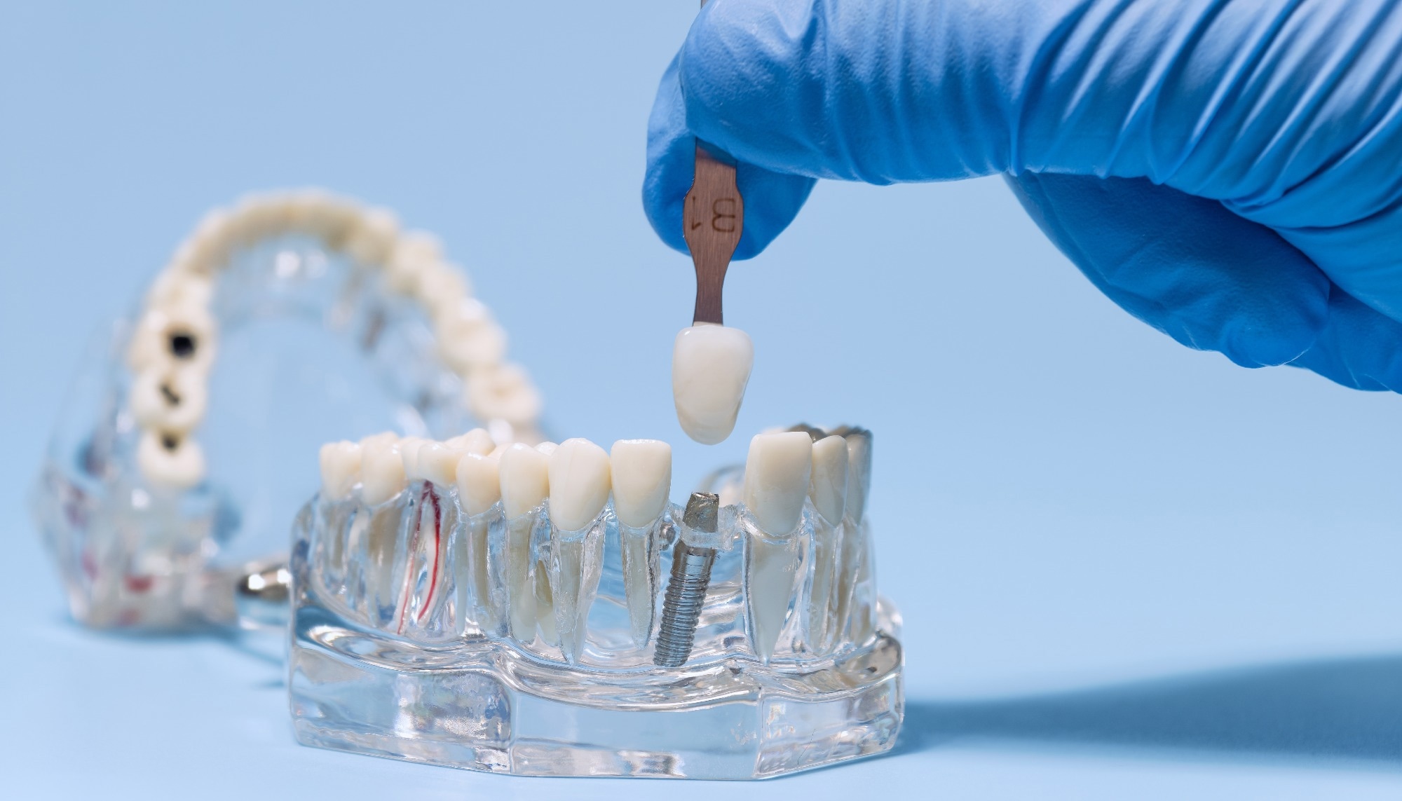 Image credit: Zakharchuk/Shutterstock.com
These models support early detection through dysbiosis sensors and may provide a foundation for future clinical applications. However, the authors emphasize that in vivo validation and incorporation of host interactions are still required before they can be translated into prevention or treatment strategies.
Oral biofilms (dental plaque) are complex bacterial communities that can shift into a dysbiotic state, promoting various oral diseases. Protected within their extracellular matrix, these bacteria resist immune defenses and antibiotics, complicating treatment. Biofilm formation poses a greater risk on endosseous implants, as implants lack innate defense mechanisms, increasing susceptibility to peri-implant diseases such as peri-implantitis and mucositis.
These diseases can rapidly lead to inflammation, bone loss, and implant failure. Therefore, early detection of dysbiosis is critical. Robust in vitro models replicating natural oral conditions are essential for developing diagnostic tools and testing preventive and therapeutic strategies.
About The Study
In the present study, researchers developed and compared in vitro multispecies oral biofilm models to examine the influence of cultivation time and species composition on the dynamics of peri-implant biofilms. They established commensal and dysbiotic models.
In the commensal model, S. oralis initially dominated but declined over time, while V. dispar and A. naeslundii stabilized at ~30% each, creating a diverse community with reduced pH. In contrast, increased pathogenic species, elevated pH, and virulence factors characterized the dysbiotic model.
The team adapted the Hannoverian Oral Multispecies Biofilm Implant Flow Chamber (HOBIC) model to reproduce peri-implantitis-associated dysbiosis. Using static and dynamic setups, they cultivated biofilms on titanium discs over 21 days under anaerobic conditions at 37°C. The models included five bacterial strains: Actinomyces naeslundii, Streptococcus oralis, Fusobacterium nucleatum, and Veillonella dispar with Porphyromonas gingivalis DSM 20709 (commensal model) or Veillonella parvula with P. gingivalis ATCC W83 (dysbiotic model).
The team standardized the inoculum to an optical density of 0.05 at 600 nm (OD600) in supplemented brain heart infusion (BHI) medium. They modified the HOBIC system by incorporating pH-sensitive flow-through cells connected to optical fibers and titanium discs immersed in an artificial saliva solution comprising mucin, lysozyme, α-amylase, and albumin. They included F. nucleatum in both models to promote interspecies interactions.
The team monitored bacterial growth and metabolism via pH and optical density measurements. They performed live/dead staining and confocal fluorescence microscopy, followed by digital image interpretation, to assess the biofilm structure and viability. They conducted real-time polymerase chain reaction (PCR) with fluorescence in situ hybridization (FISH) to determine species distribution. Additionally, they evaluated gingipain levels in the flow chamber supernatants by a P. gingivalis-specific enzyme-linked immunosorbent assay (ELISA) to assess the activity of virulence factors.
Results
Biofilm development was strongly shaped by bacterial species composition, with cultivation conditions playing a lesser, but still measurable role. In both static and dynamic systems, dysbiotic shifts occurred through intrinsic bacterial interactions under flow conditions, without the application of external stimuli.
In the commensal model, microbial composition diversified over time, accompanied by reduced pH values (<6.3). By contrast, the dysbiotic model exhibited greater overall growth, higher pH values (up to 6.9), and increased expression of virulence factors. Biofilm volume in dysbiotic models increased sharply after the first day and stabilized within six days (HOBIC) to 15 days (static), while in commensal biofilms, it initially decreased and then re-established. Biofilm viability declined over time under most conditions, reflecting maturation and nutrient limitations. The amount of gingipain protein significantly increased over time within the dysbiotic model only.
In commensal models, S. oralis initially dominated (70%) but declined over time, while V. dispar and A. naeslundii stabilized at 30% each. F. nucleatum and P. gingivalis remained nearly undetectable. In dysbiotic models, V. parvula initially dominated (95%) but decreased under flow, while A. naeslundii, F. nucleatum, and P. gingivalis increased (15–30% each). Static dysbiotic biofilms maintained V. parvula dominance (50–60%) alongside higher P. gingivalis and F. nucleatum.
Commensal biofilms acidified the medium through S. oralis fermentation, whereas dysbiotic biofilms, dominated by Veillonella, generated less acidic metabolites, maintaining a higher pH. These conditions favored P. gingivalis proliferation. Gingipain levels significantly increased only in dysbiotic biofilms.
Metabolic analysis suggested interspecies nutrient sharing. Commensals produced metabolites such as lactate, peptides, and vitamins, which supported the growth of F. nucleatum and P. gingivalis. The study noted that V. parvula may contribute to thiamine biosynthesis but emphasized that strain-level effects and complex metabolic interactions likely play a larger role in driving dysbiosis.
Conclusions
The study established advanced in vitro multispecies biofilm models, demonstrating that bacterial species selection, particularly V. parvula and the clinical P. gingivalis strain W83, was the primary driver of dysbiotic shifts, with cultivation conditions playing a secondary but relevant role.
The adapted HOBIC flow chamber effectively reproduced peri-implantitis-associated dysbiosis, providing a valuable platform for investigating disease mechanisms and developing early detection tools such as dysbiosis sensors.
However, the authors caution that these simplified in vitro models do not yet account for immune responses, host tissues, or the oral microbiome's full diversity; therefore, in vivo validation is essential before clinical application.
Journal Reference
Heine, N. et al. (2025). Influence of Species Composition and Cultivation Conditions on Peri-Implant Biofilm Dysbiosis in Vitro. Frontiers in Oral Health, 6, 1649419. DOI: 10.3389/froh.2025.1649419. https://www.frontiersin.org/journals/oral-health/articles/10.3389/froh.2025.1649419/full
 Type I CRISPR Systems Regulate Embedded Innate Immunity Genes in Neisseria