Amino acids play a dual role in cellular metabolism. They are the major building blocks in protein synthesis while also serving as intermediary metabolites to drive biosynthetic reactions. Accurate quantification of L-amino acids in purified samples or body fluids can provide crucial information for basic research and diagnostic purposes.
Cellular Roles of Amino Acids in Cancer and Neurobiology
The metabolism of cancer cells, which are recognized for their metabolic abnormalities, is considerably altered. Warburg effect is one such abnormality in which increased glycolytic activity takes place even in the presence of oxygen. The high rate of aerobic glycolysis enables the steady growth and survival of cancer cells.
Biosynthetic pathways and cancer cells mostly consume amino acids as nutrients and, as a result, amino acids are always studied extensively for different subtypes of cancer. It can be seen that components sensing the adequacy of amino acids in cancer tissues exhibit a slight modification. This results in cells with the mechanistic target of rapamycin (mTOR) that regulates autophagy and the synthesis of proteins.
These processes alter the metabolism of proliferative cells to promote the biochemical pathways for the accumulation of biomass; hence, such alterations in the tumor cell metabolism are believed to be cancer characteristics.
Researchers have demonstrated that the levels of metabolites and metabolic activities can help differentiate tumor tissues from normal tissues. Hence, any data pertaining to this differentiation can be carefully used for detecting and treating cancer. Having a deeper understanding of the altered metabolism in malignant cells and tissues has a higher potential for synergy with improved medical treatments for this type of disease.
For example, researchers detected high levels of the nutrient asparagine in rapidly growing cancer cells which ultimately led to the discovery of L-asparaginase treatment for leukemia.
In addition, the numerous affinities of the uptake of glucose in certain tumors presented new opportunities for developing the 18fluoro-2-deoxyglucose imaging agent for positron emission tomography (PET). This outcome motivated many researchers to examine the metabolism of tumor glucose.
In neurobiology, it is a well-known fact that the brain contains higher concentrations of amino acids when compared to other body tissues. Neurons react to amino acid neurotransmitters, such as gamma-aminobutyric acid (GABA), glutamate, glycine (major inhibitory neurotransmitters), aspartate, and N-acetyl aspartate (excitatory neurotransmitters).
Ionotropic glutamate receptors like N-methyl-D-aspartate (NMDA) and α-amino-3-hydroxy-5-methyl-4-isoxazolepropionic acid receptor (AMPA) play a role in the regulation of glutamatergic neurotransmission. Amino acids like tryptophan and phenylalanine also have an important role to play in the production of serotonin and dopamine, and in sustaining sleep cycles.
Therefore, amino acids are highly crucial components to ensure a healthy mental state. Recent analyses have shown that one of the most common changes observed in cancer is caused by the deregulation of both arms of these amino acids.
Amino Acid-Based Assays
Assay kits meant for detecting L-amino acids are important tools for acquiring crucial data in studies.
Glutamine
Glutamine (Gln) is vital for many biological processes such as the regulation of acid balance in mammalian kidneys, cell growth, and protein synthesis (refer to Figure 1).
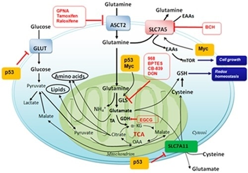
Figure 1. Glutamine and the metabolism of other amino acids as targets for cancer therapy. (Int J Mol Sci 2015;16:22830–22855; doi:10.3390/ijms160922830). Image Credit: BioVision Incorporated.
Glutamine is the major source of nitrogen for cells, to produce nucleotides and hexosamines. It even plays a role in cancer signaling, energy production, and redox homeostasis.
Specific cancer cell lines have been observed to display an affinity towards glutamine. Tumors addicted to glutamine are likely to present oncogenic expression of the Myc gene. This Myc gene codes for a transcription factor that encourages the expression of glutamine transporters and metabolic enzymes for biosynthesis. The result is cells that experience aerobic glycolysis ensuring a continuous influx of glucose.
All these facts make glutamine detection an interesting target for upcoming therapeutic and diagnostic options for cancer. Glutamine plays an important role in triggering mTOR complex 1 and maintaining the activity of TOR kinase. These activities integrate the metabolism by detecting the nutrient levels and controlling the production levels of other amino acids, along with the biosynthesis of lipids.
A commercial glutamine colorimetric assay kit is now available. This kit has the potential to detect biologically relevant glutamine concentrations in various biological fluids and tissues, which accordingly help in expediting the discovery process.
Glycine
Glycine (Gly) is a crucial component of body proteins that are responsible for building tissues that form joints, muscles, and organs. It is abundantly present in collagen and is the second most abundant amino acid in human proteins and enzymes.
Glycine functions as an inhibitory neurotransmitter in the central nervous system (CNS) and thus plays a significant role. This neurotransmitter processes both sensory and motor information, enabling audition and movement mediated by a glycine receptor that is sensitive to strychnine.
At times, glycine is jointly released with GABA—the major inhibitory amino acid neurotransmitter. It reinforces the glutamate action at NMDA receptors and thus regulates the excitatory neurotransmission.
Glycine is used for treating schizophrenia, stroke, benign prostatic hyperplasia (BPH), and other various disorders. These applications of glycine have not led to any evident adverse events.
Measuring the endogenous levels of glycine or glycine in in-vivo samples is slightly difficult. Currently, no accurate method is available for high-throughput screening of the concentrations of glycine in samples. The standard protocols for glycine quantitation are nuclear magnetic resonance (NMR), high-performance liquid chromatography (HPLC), mass spectrometry (MS), and immunoassay techniques.
However, these procedures are not suitable for compound screening or for a fast-paced screening of a large number of samples. Glycine detection kits that are available in the market are useful for high-throughput screening to quantify the concentrations of glycine in numerous biological samples. These kits provide simple, sensitive, and reproducible methods for the detection of glycine, which are relevant for in-vitro serum screening of small-molecule activators or inhibitors.
These kinds of assays have shown that there is a significant reduction of glycine in patients suffering from depression and schizophrenia; The sample collected from a patient suffering from Alzheimer’s disease had a higher concentration of glycine (refer to Figure 2). Similar trends were also shown by the latest studies.
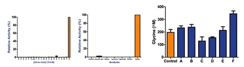
Figure 2. (A and B) Commercial glycine detection kit results showing that metabolites and other amino acids do not interfere with glycine detection. (C) The assay used for research demonstrating a decrease of glycine in patients suffering depression and schizophrenia and increased concentration of glycine in Alzheimer’s disease patients. Glycine concentration was evaluated using an in vitro commercial glycine assay kit in human serum samples from patients diagnosed with various neuropsychiatric conditions. Control: Control; A: Multiple Sclerosis; B: Parkinson’s Disease; C: Major Depression; D: Schizophrenia; E: Amyotrophic Lateral Sclerosis; F: Alzheimer’s Disease. All patients were male Caucasian and age ranges from 53 to 60 years old. Image Credit: BioVision Incorporated.
L-Tryptophan
L-Tryptophan (TRP) is one of the eight amino acids that are essential to the human body. It plays a crucial role in the endogenous synthesis of melatonin, serotonin, protein, niacin, tryptamine, kynurenine, nicotinamide adenine dinucleotide (NAD), and nicotinamide adenine dinucleotide phosphate (NADP). Kynurenic acid produced from kynurenine is a glutamate receptor antagonist.
Serotonin synthesis is one of the most important tryptophan pathways (refer to Figure 3). Tryptophan decarboxylation leads to the formation of tryptamine, which is a vital serotonin neuromodulator. Melatonin is a hormone that is produced from the tryptophan–serotonin pathway. This pathway regulates biological rhythms, like diurnal rhythms that stabilize the hormonal and cardiac systems.
Tryptophan has an effect on other CNS molecules and neurotransmitters like beta-endorphin, dopamine, and norepinephrine, thus displaying a wide range of neurophysiological effects in the human body.
Distinctive fluorometric properties are chemically presented by the TRP side chain (indole). TRP is the only amino acid that can be present in dual forms in the blood—that is, bound and free forms.
Differences in the concentrations of tryptophan are directly connected to numerous physiological and behavioral processes, like motion, sleep, memory, sickness, schizophrenia, depression, and bipolar disorders.
In vitro detection of TRP that is available in the market provides a simple, sensitive, and high-throughput adaptable assay with the potential to identify the concentration of tryptophan in biological fluids, including free and bound tryptophan in serum.
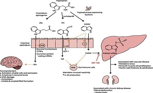
Figure 3. Tryptophan functions in the human body. (Zhang LS, Davies SS via Wikimedia Commons). Image Credit: BioVision Incorporated.
Conclusion
Amino acids are essential constituents of several cellular and structural proteins. Such building blocks can prove to be useful tools for a wide range of studies.
About BioVision Incorporated
BioVision, Inc., is a privately held Life Science company headquartered in the beautiful San Francisco Bay Area.
BioVision develops and offers a wide variety of products including assay kits, antibodies, recombinant proteins & enzymes, and other innovative research tools for studying Apoptosis, Metabolism, Cell Proliferation, Cellular Stress, Cell Damage and Repair, Diabetes, Obesity and Metabolic Syndrome, Stem Cell Biology, Gene Regulation, Signal Transduction, etc. BioVision's products are currently being sold in more than 60 countries worldwide.
Sponsored Content Policy: News-Medical.net publishes articles and related content that may be derived from sources where we have existing commercial relationships, provided such content adds value to the core editorial ethos of News-Medical.Net which is to educate and inform site visitors interested in medical research, science, medical devices and treatments.