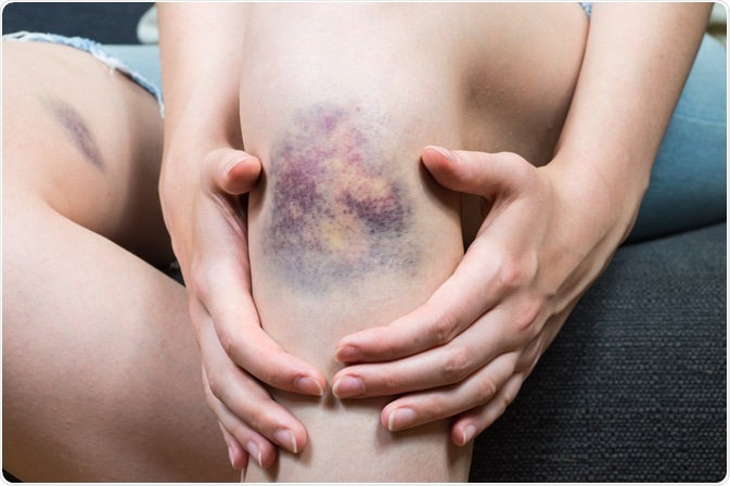
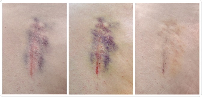
Medical examiners often encounter questions about the duration of injuries based on the bruises encountered on a human body. Visual determination through photos or direct examination is usually used as a method to assess a time estimation on the skin hematomas.

Image Credit: Aleksey Boyko/Shutterstock.com
However, the disadvantages of relying on visual inspections include the lighting of the room or where the photo was taken as well as the quality of the photograph.
Bruised areas on the human body usually undergo color development within a set period and studies have shown that a spectroscopy technique can be a useful technique to observe skin modifications, such as discoloration and texture.
Reflectance spectroscopy
Reflectance spectroscopy is commonly used as a diagnostic tool by giving insights regarding the structure of tissues through their absorption spectrum. Specifically, the hemoglobin concentration (Hb) of the tissue as well as the heme oxygenase system.
For instance, the oxygenated hemoglobin would reflect the color red when it absorbs the blue light as a result of when white light shines on the object. While the hemoglobin that is deoxygenated may give off a more pigmented blue light color after absorbing a red light with a higher level when the white light illuminates it.
This technique is advantageous for its ability to give measurements without touching the sample, hence minimum contamination and preservation of the sample are maintained. The reflectance spectroscopy system consists of a light source with a high enough brightness so that the background noise can be minimized by making the reflected signal much stronger.
Nonetheless, the light source power must not surpass the maximum power set by the American National Standards Institute (ANSI) when performing the analysis in-vivo to avoid damaging the tissue studied.
The fiber optic probe component collects the signal that is reflected in the tissue from the light being excited. The geometry of the fiber probe controls the depth of tissue penetration. The signal detected is then split into each wavelength by the spectrometer and their intensities are detected by using a charge-coupled device as the detector.
Absorption and scattering on human skin
Light can either be transmitted, scattered, reflected, or absorbed after photons are propagated on the biological tissue, depending on the tissue's optical properties and its wavelength.
The photons can only be absorbed if their energy matches the energy of the molecules and the energy state of the molecule that absorbed this photon is then exited. While scattering is caused by the change in the refractive index that deflects the electromagnetic radiation or particles, where the size of the particle greatly impacts how the light is scattered.
These properties can be applied to the human skin, as the skin contains chromophores such as bilirubin, hemoglobin, and melanin that is responsible for the color appearance. The visual characteristics of the skin are caused by the properties of tissue scattering or absorption.
Color changes in bruised areas
Color changes occurring with time on the bruise areas are caused by the transport of hemoglobin into the dermis as well as the breakdown products. This color development can be monitored by reflectance spectroscopy.
Randeberg, et. al use the Fick's law of diffusion as well as Darcy's law to study these occurrences as a function of time from the experimental data obtained using the technique. The spectra are set to the wavelength range of 400 - 850 nm on the normal and bruised skin.
One of these hemoglobin breakdown products is bilirubin, which causes the color yellow on the bruised area. As discussed, the color of bilirubin absorbs at the wavelength of approximately 460 nm, where the complementary of this wavelength produces the yellow color.
Bohnert et al. uses a reflectance spectroscopy technique to examine post-mortem bruises and studied that recent and mild contusions usually generate the color red, while more severe bruises appear in the shade of blue.
Chemical signals released as a result of trauma send macrophages to the injured site, as they are responsible for breaking down the hemoglobin and the hemoglobin is catabolized through the heme oxygenase system.
Heme oxygenase breaks the heme closed ring structure so that biliverdin, the green pigment in human skin, can be formed. Nonetheless, biliverdin only exists in a short amount of time for its high reductase activity, and bilirubin is then gradually removed from the bruised area. After trauma, the chromophore hemosiderin appears in the color brown for a few months as a result of heme breakdown products.

Image Credit: Volodymyr Nikitenko/Shutterstock.com
Limitations of spectroscopy to study aging bruises
Reflectance spectroscopy utilizes the point-probe component that makes the reflected light intensity as well as the color measurement more convenient, however, the tool can only analyze a limited number of tissues. The limitation of this component makes the analysis time consuming as only a portion of the tissue sample can be diagnosed one at a time.
Furthermore, there needs to be more samples of bruises to be examined to validate the results of this diagnosis using reflectance spectroscopy. Once these limitations are overcome, reflectance spectroscopy can be a useful technique in more medical practices by providing a more accurate analysis of a larger tissue area.
References
- Mao, S., Fu, F., Dong, X. and Wang, Z. (2011). Supplementary Pathway for Vitality of Wounds and Wound Age Estimation in Bruises Using the Electric Impedance Spectroscopy Technique. Journal of Forensic Sciences, [online] 56(4), pp.925-929.
- Wallace, M., Wax, A., Roberts, D. and Graf, R. (2009). Reflectance Spectroscopy. Gastrointestinal Endoscopy Clinics of North America, [online] 19(2), pp.233-242. Available at: https://www.ncbi.nlm.nih.gov/pmc/articles/PMC2841958 [Accessed 17 August 2020].
- Randeberg, L. (2020). Diagnostic Applications Of Diffuse Reflectance Spectroscopy. PhD. Norwegian University of Science and Technology. Available from: NTNU Open [Accessed 17 August 2020]
- Randeberg, L., Haugen, O., Haaverstad, R. and Svaasand, L. (2006). A novel approach to age determination of traumatic injuries by reflectance spectroscopy. Lasers in Surgery and Medicine, [online] 38(4), pp. 277-289.
Further Reading
Last Updated: Oct 1, 2020