Electron Microscopy provides key structural information on a wide range of specimens, from material characterization to biomedical investigation. Technological advances are contributing to the continuous global market growth, together with increasing research-based activities, particularly in the life sciences and the nano- and biotechnology sectors.
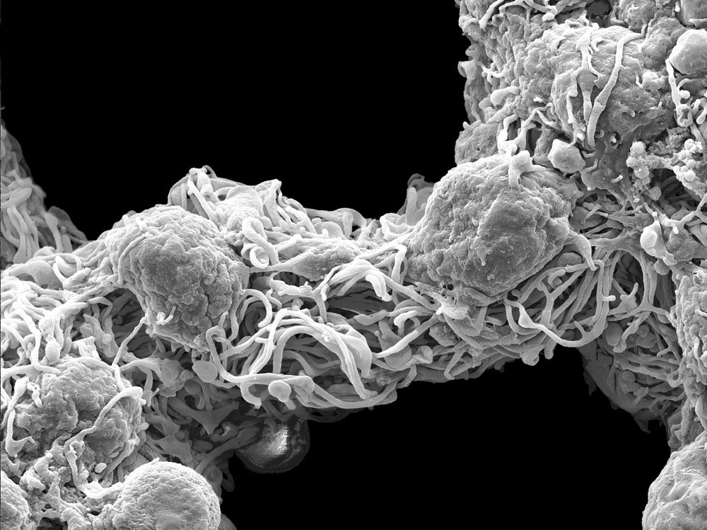
Image Credit: thomaslabriekl/Shutterstock.com
Application Areas of Electron Microscopy
Electron microscopy (EM) comprises a series of techniques for obtaining high-resolution images of both organic and inorganic specimens. EM uses electrons as the source of illuminating radiation and allows to study samples at the sub-nanometer scale, investigating their shape, crystal structure, chemistry, as well as their electric and magnetic properties.
The most common types of EM are Transmission Electron Microscopy (TEM) – which detects electrons that pass through very thin samples – and Scanning Electron Microscopy (SEM), where the detection of electrons reflected off the surface of the sample determines the image. Cryo-EM is a groundbreaking variation of TEM where the native structure of specimens is preserved by quickly cooling them to cryogenic temperatures, preventing water molecules from crystallizing.
Major applications of EM are found in the material sciences and nanotechnology sectors, for instance, the characterization and study of the structure of semiconductors and next-generation electronics materials. Industrial applications involve quality control of products, with the analysis of defects and faults. Other applications are in forensics, healthcare, or food industry.
Over the last few years, significant progress has been made in the life sciences, where EM is applied in clinical trials or disease diagnosis and can provide information on cell functions. The technique is now extensively used to investigate the structure of tissues, cells, and macromolecular complexes.
EM is extensively used in the observation and discovery of new viruses. In the late 1940s, the differences between smallpox and chickenpox viruses were demonstrated by EM. The technique provided the first image of poliovirus (1952) and contributed to the discovery of norovirus (1972, Siemens Elmiskop 1A electron microscope).
Currently, EM is used in viral diagnostics (i.e., for detecting new and unusual outbreaks) and is a powerful tool in hospital settings. For instance, urine analysis via EM (LEO EM-910, by Zeiss) can indicate polyomavirus infection in kidney transplant patients and distinguish it from other virus infections.
Recent Developments in Electron Microscopy
In the last decade, the application of EM (particularly SEM) in the biological sciences and biomedical research has attracted much interest from scientists worldwide. It has been possible to elucidate the structure of bio-specimens with an extremely high level of detail.
A particularly hot topic is the application of SEM in neuroscience. Scientists were able to conduct a three-dimensional (3D) reconstruction of neural tissue sample images in mice, allowing them to study neurons and their connections with unprecedented detail.
Serial block-face scanning electron microscopy (SBEM) was used to measure synaptic size during the wake/sleep cycle of thousands of synapses in two regions of the mouse cortex. Images were obtained using a Zeiss SIGMA VP field emission scanning electron microscope equipped with 3View® technology.
Thanks to advances in sample processing and the introduction of new-generation cameras, applications of cryo-EM have seen rapid intensification, with consequent growth of the cryo-EM market.
A major contribution of such growth is cryo-EM's unique ability to elucidate biomolecules' high-resolution structures in situ. The technique has been so revolutionary that in 2017 three scientists, who pioneered the field of cryo-EM, were awarded the Nobel Prize in Chemistry.
Recently, cryo-EM has been extensively used in the structural characterization of SARS-CoV-2 viral proteins, which can be attractive targets for vaccine development and new drug formulations to combat viral infections. One example is the explanation of the mechanism of SARS-CoV-2 RNA polymerase inhibition by Remdesivir (an RNA nucleotide analog that binds to the replicating RNA blocking viral replication). The study was performed on a Titan Krios equipped with a Gatan K2 Summit direct electron detector.
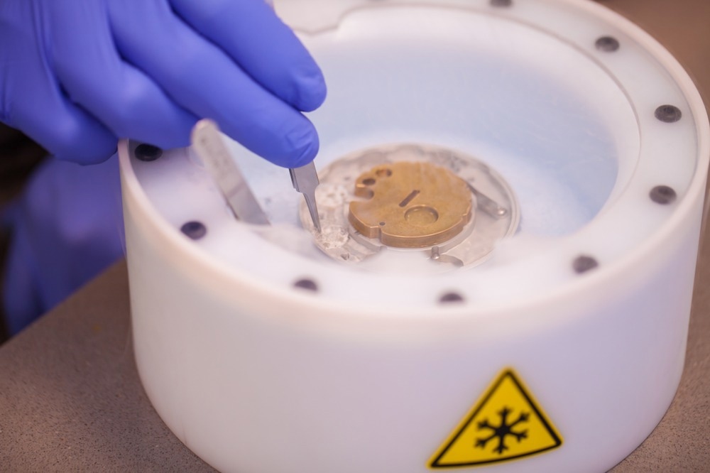
Image Credit: PolakPhoto/Shutterstock.com
Current Global Market of Electron Microscopy
In 2021 the global market of Electron Microscopy was valued at $3.7 billion, and by 2031 it is expected to reach $8.3 billion. The compound annual growth rate (CAGR) – the mean annual growth rate over a specified period – from 2022 to 2031 is projected at 8.5%.
The biggest revenue contributor to the EM market comes from SEM. The leading area is Asia Pacific, with more than 36% of the revenue share in 2020. This is due to the rapid increase in applications in semiconductors, pharmaceuticals, and nanotechnology.
North America is the second-largest regional market for SEM, with a revenue share of over 34%. Valued more than $1 billion in 2022, the SEM market is estimated to expand due to a surge in demand for pharmaceutical research. This will be especially boosted by the demand for SEM in research and academic institutes and the high number of clinical trials performed in the region.
The global market of EM is also witnessing impressive growth in the cryo-EM segment. The cryo-EM market was, in fact, over $725 million in 2021 and is expected to exceed $1.5 billion by 2028, with a forecasted CAGR of 11.2%.
The presence of state-of-the-art diagnostic centers and ongoing clinical trials in North America boost the cryo-EM market. Nevertheless, the rapid technological advancements and increased healthcare expenditure in Asia are projecting the region to have the highest CAGR.
There are many companies worldwide that are at the forefront of electron microscopy. Some key players include Amgen, Bruker, Hitachi, JEOL, Nikon Instruments, Oxford Instruments, Thermo Fisher Scientific, and Zeiss.
Late in 2020, Zeiss launched three new products in the field emission scanning electron microscope (FE-SEM) family – the ZEISS Gemini SEM 360, 460, and 560. Similarly, in August 2020, JEOL announced the launch of JSM-IT700HR, a new scanning electron microscope, to strengthen its growth. In October 2021, Amgen and the University of Southern California (USC) teamed up to deliver two cutting-edge microscopes, the Thermo Scientific Krios and Glacios Cryo-TEMs.
Considerable market growth in the upcoming years will be driven not only by the increasing number of laboratories working on semiconductors and material sciences but also by the increasing use of EM to study surface morphology and chemical analysis. In addition, the increased investments in R&D-related EM applications to viruses and chronic diseases will offer significant growth opportunities for the market.
Future Directions for Electron Microscopy
The field of EM is constantly evolving. As David Muller (applied physicist, Cornell University in Ithaca, New York) commented in an editorial on Nature, "I don't think anyone is standing still. Everyone's thinking about what you want to build next".
Many challenges remain, with efforts dedicated to improving the resolution of electron microscopes and obtaining images with greater details. Advances may be driven by improvements in the electron beams themselves, with monochromators that can narrow the range of energies for electrons that reach the sample.
Future developments focus on determining the functionality of materials at atomic or molecular resolution. The complex 3D arrangements of atoms (and defects) determine the properties of materials. Consequently, future opportunities for EM lie in the better 3D imaging of materials, especially on defects, dopants, and impurities, along with their chemical identification to correlate them with the material's behavior.
While applications of EM continue to be strongly linked to materials characterization, the area that seems to be driving the technological advancements is related to cryo-EM. A factor that could further enhance the power of cryo-EM is the ability to capture transient intermediate states in macromolecules (i.e., short-lived conformations of proteins) and their complexes.
This could reveal unprecedented insights into the mechanisms and functions of proteins. However, current methods fail to do so because they are too slow; therefore, a lot of research is focused on solving the issue.
Moreover, other areas of development, such as automated pipelines for data processing, and the availability of low-cost, user-friendly microscopes, are key directions to pursue to enhance the accessibility and usage of EM.
Final Thoughts
Over the last decades, EM consolidated its role within the scientific community, contributing to cutting-edge research. Technology advancements are leading to an expansion of applications in a wide range of sectors, with industry and research organizations working together to overcome technical barriers. As a result, the global electron microscopy market is projected towards rapid growth in the upcoming years.
Sources:
- Goldsmith, C. S. & Miller, S. E. (2009). Modern uses of electron microscopy for detection of viruses. Clin Microbiol Rev, 22, 552-63.10.1128/CMR.00027-09
- De Vivo, L., Bellesi, M., Marshall, W., Bushong, E. A., Ellisman, M. H., Tononi, G. & Cirelli, C. (2017). Ultrastructural evidence for synaptic scaling across the wake/sleep cycle. Science, 355, 507-510.doi:10.1126/science.aah5982
- Assaiya, A., Burada, A. P., Dhingra, S. & Kumar, J. (2021). An overview of the recent advances in cryo-electron microscopy for life sciences. Emerg Top Life Sci, 5, 151-168.10.1042/ETLS20200295
- Yin, W., Mao, C., Luan, X., Shen, D. D., Shen, Q., Su, H., Wang, X., Zhou, F., Zhao, W., Gao, M., Chang, S., Xie, Y. C., Tian, G., Jiang, H. W., Tao, S. C., Shen, J., Jiang, Y., Jiang, H., Xu, Y., Zhang, S., Zhang, Y. & Xu, H. E. (2020). Structural basis for inhibition of the RNA-dependent RNA polymerase from SARS-CoV-2 by remdesivir. Science, 368, 1499-1504.10.1126/science.abc1560
- Courtland, R. (2018). The microscope revolution that's sweeping through materials science. Nature, 563, 462-464.https://doi.org/10.1038/d41586-018-07448-0
Further Reading
Last Updated: Oct 28, 2022