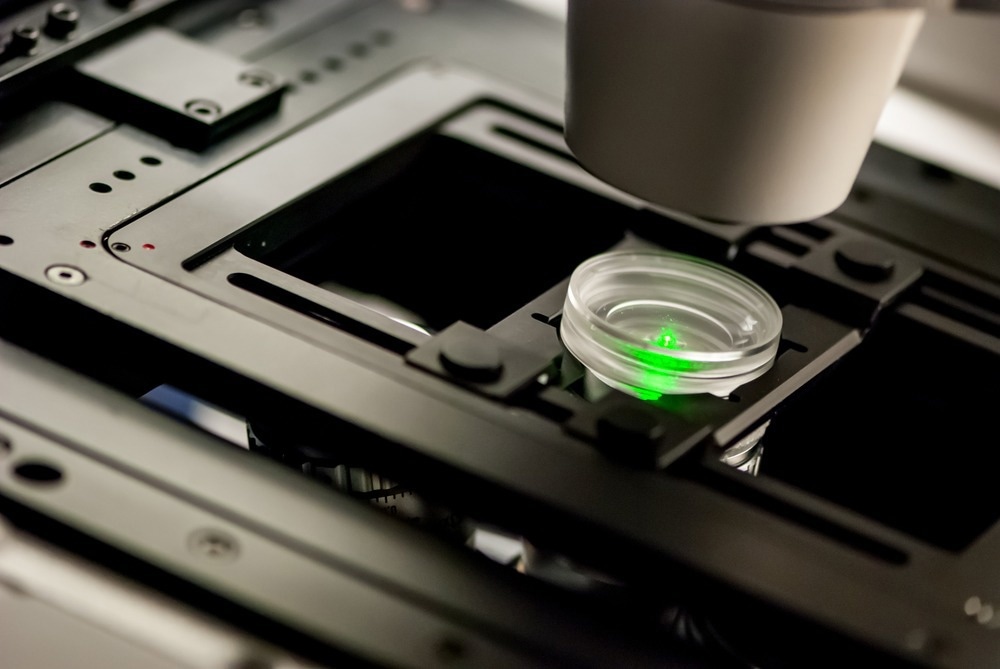
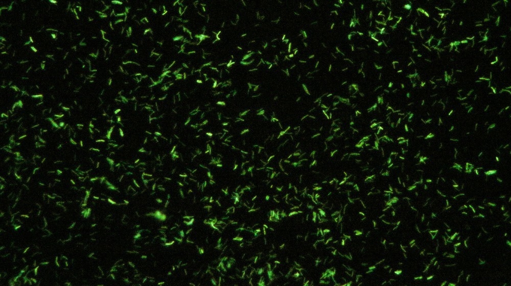
Fluorescence microscopy is a workhouse technique in the life sciences for tissue analysis, cell structure visualization, and the study of biological processes and interactions.1 More specifically for genetics research, typical uses of fluorescence microscopy include performing high throughput screening processes and studying the structure of genetic material.
 Image Credit: Micha Weber/Shutterstock.com
What is Fluorescence Microscopy?
Fluorescence microscopy is an optical microscopy technique that works by detecting the emitted fluorescence from a sample. Normally, samples are labeled with a fluorescent tag known as a fluorophore – a chemical species that emits light on being photoexcited with a light source such as a laser.
Most standard optical microscopies illuminate a sample with white light and look at how much light is absorbed by different regions of the sample. The advantage of fluorescent techniques, particularly with advanced genetic modification methods, is that fluorophores can be used to label very specific parts of a genetic sequence or cell structure.2 When dealing with large or more complex samples, certain structures or regions can be selectively imaged – helping to deconvolute features that might be difficult to distinguish with standard transmission-based optical methods.
With a wide range of fluorophores now available, fluorescence microscopy can be a highly efficient technique for imaging multiple structures in a single experiment using optical filters.
If several cell sections are labeled with fluorophores that emit at different wavelengths, filters or selective excitation wavelengths can be used to only visualize one fluorescence wavelength at a time.3 The information from different fluorophores can then be correlated to gain greater insights into the structure and, in particular, dynamical processes happening in the biological system.4
The development of readily available commercial solid-state lasers and microscopy platforms has helped the uptake of fluorescence microscopy in the life sciences considerably. Many companies offer all-in-one systems of the light source and microscope as well as acquisition software that can not just help with capturing images but with automated processes such as cell counting, cell structure labeling and Z-stacking – where images are taken at different focal distances and then summed together to try and reconstruct a 3D, rather than 2D image.
Some examples of fluorescence microscopy platforms include KEYENCE’s instruments as well as long-established optics experts Leica, who offer a number of widefield fluorescence microscopy instruments. ThermoFischer Scientific offers all-in-one platforms like the EVOS M5000 imaging system that is designed specifically for use by cell imaging labs and even includes computer hardware such as an integrated monitor.
Greater automation capabilities also exist for microscope control and experiment design, with features such as automated focus finding helping improve the throughput of fluorescence microscopy platforms. Improved image quality and compatibility with advanced imaging stages, such as incubators, has enabled live cell imaging of cell growth and development.
How Can Fluorescence Microscopy Be Used in Genetics Research?
One example of a genetics study that makes use of fluorescence microscopy is the use of time-lapse fluorescence microscopy to study gene regulation.5 Time-lapse microscopy involves taking multiple images over a given time period to see how a process evolves as a function of time.
Time-lapse microscopy is very powerful in genetics research because the movement of the fluorescent signals can be used to study gene dynamics and movement, and the use of particular fluorophore tags can also be used to look at properties such as inheritance. Depending on how the tag is created and attached to the species of interest, the tag may be transferred during processes such as cell differentiation and so the tag can be tracked as the biological process unfolds.
One of the advantages of using fluorescence microscopy for gene regulation studies is that it is a relatively non-intrusive method – particularly for fluorescent proteins that do not require the introduction of a fluorophore tag for imaging. Automation of processes, such as autofocusing, has also helped improve the quality of data that can be obtained through time-lapse methods.

Image Credit: vchal/Shutterstock.com
Challenges of Using Fluorescence Microscopy for Genetic Applications
The ability to introduce genetic mutations into proteins to introduce fluorescence and the availability of larger numbers of fluorescence tags have helped make fluorescence microscopy compatible with a larger number of chemical and biological systems.
However, finding good sample preparation procedures with some tags can be difficult and needing a tagged species can mean some small perturbations are introduced into the system.
As the fluorescence yield is dependent on the excitation intensity, it can seem reasonable to use higher average laser powers to perform fluorescence microscopy measurements. However, photodamage is a significant issue that limits the overall acquisition time of an image.
The problem of photodamage and photodegradation is generally worse at higher laser powers and so it can be challenging to measure very weak fluorescence signals. This is also a problem for several electron microscopy approaches that generally offer better spatial resolution than optical microscopies but at the cost of more expensive instrumentation and greater problems with sample damage.
Fluorescent microscopy is a robust and versatile technique offering enhanced selectivity over standard confocal approaches. The development of superresolution methods is helping overcome the relatively large resolution limit set by the wavelength of visible light. More and more fluorophores are becoming available, making it possible to measure a greater number of systems.
Sources:
- Drummen, G. P. C. (2012). Fluorescent probes and fluorescence (microscopy) techniques-illuminating biological and biomedical research. Molecules, 17(12), pp. 14067–14090. doi.org/10.3390/molecules171214067
- Renz, M. (2013). Fluorescence microscopy-A historical and technical perspective. Cytometry Part A, 83(9), pp. 767–779. doi.org/10.1002/cyto.a.22295
- Hickey, S. M., et al. (2022). Fluorescence microscopy—an outline of hardware, biological handling, and fluorophore considerations. Cells, 11(1). doi.org/10.3390/cells11010035
- Yu, L., et al. (2021). A Comprehensive Review of Fluorescence Correlation Spectroscopy. Frontiers in Physics, 9, pp. 1–21. doi.org/10.3389/fphy.2021.644450
- Zou, F., & Bai, L. (2019). Using time-lapse fluorescence microscopy to study gene regulation. Methods, 159–160, pp. 138–145. doi.org/10.1016/j.ymeth.2018.12.010
Further Reading
Last Updated: Sep 17, 2024