Subcellular imaging can be a very powerful tool for understanding disease at a cellular and molecular level. This can enable labeling and imaging of biological systems that can aid in visualizing and quantifying nutrients, toxic elements, and drugs in single cells.
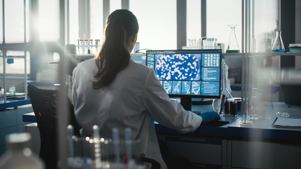
Image Credit: Gorodenkoff/Shutterstock.com
The significance of this process includes the impact of disease pathways at the subcellular level and the regulation of these pathways by drugs for disease treatment and prevention.
Drug Discovery
The treatment of disease is heavily interlinked with drug discovery and development, initiated due to unmet clinical needs. The development of drugs from the initial idea stage, research development, to the finished product can take between 12 to 15 years and cost over $1 billion.
The process of drug discovery can include: (i) basic research, (ii) lead discovery, (iii) preclinical discovery, (iv) clinical development, and (v) Food and Drug Administration (FDA) filing. This process entails identifying and selecting the target compound, the development of the drug candidate, progression into preclinical trials, and if successful, clinical development to gain FDA approval for the marketing of the medicine.
Screening assays are a large component of the drug discovery process, and this can comprise target validation, compound screening, secondary assays, and in vivo analysis.
Identification of Drug Targets and Challenges
Target identification is a significant portion of drug discovery as it can ensure the drugs being developed are effective and safe – two predominant causes of drug failure within the clinic. A 'good' target, which can include a wide variety of biological entities, such as proteins, is required to elicit a biological response that can be measured in vivo and in vitro. Subcellular drug imaging can ensure effective drug targets are chosen.
Phenotypic screening can identify disease targets and molecules that result in the desired phenotype associated with a therapeutic effect. This approach has been established in both an academic and industry setting to discover new drug targets and pathways. The analysis of drug targets and pathways is significant for research into drug development as the interaction of drugs can be assessed for their efficacy in intervening with the disease pathways for use as a potential treatment.
However, this critical interaction between small molecule therapeutic drugs and their protein targets can be poorly understood, impacting the drug's efficacy. This interaction may not be able to be visualized in live cells or even entire organisms due to unavailable methods that can be used directly to measure the engagement of drugs and their targets within a biological setting.
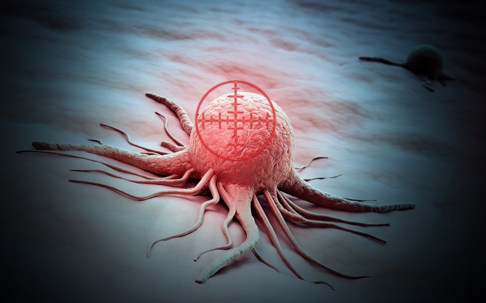
Image Credit: Mopic/Shutterstock.com
Imaging Techniques
However, recent imaging advancements have enabled pharmaceuticals to produce fluorescent drugs, prodrugs, and probes directed at targets. These drugs have predominantly been used for in vitro testing, while some have been used in vivo for imaging drug distribution and detecting tumor cells.
Fluorescence polarization (FP) has been hypothesized to be used to accurately measure drug binding in both in vivo and in vitro testing through multiphoton microscopy. This method can provide insight into the environment of the excited fluorescent molecule, which can be used to measure the number of interactions between the fluorescent molecule and molecular drug interactions.
Another potential bioimaging method is mass spectrometry imaging, which can be used for visualizing subcellular domains to investigate the organization of different biomolecules, such as for drug and medical research.
This imaging strategy has been said to be more potentially powerful with the application of quantifying biomolecules at a nanoscale, which can be possible with nanoscale secondary ion mass spectrometry (nanoSIMS). This method consists of benefits such as using non-toxic labels as well as having high spatial resolution down to 50 nm and can be advantageous when imaging drug interactions at a cellular level.
With nanotechnology becoming more interlinked with other fields and applications, using nanoscale imaging for drug development can be a useful method of understanding cell interactions. This is due to the natural interaction of cells and biological systems with drugs produced using nanocarriers. Therefore, the analysis of nanoscale drug formulations using subcellular drug imaging can hold a significant role in understanding the level of efficacy of these drugs and how they can be made with a higher level of targeting for a specific disease and pathway, as well as ensuring safety within patients.
Future Outlook
Subcellular drug imaging may be revolutionary for medicine and pharmaceutical industries as the advanced analysis of drugs in a biological setting can demonstrate their level of efficacy and safety.
This is significant for drug discovery and development as incorporating high-level drug imaging may enable the length of time for drug development to be more optimistic, as there would be a higher probability of approval from the FDA.
Continue reading about imaging techniques in drug discovery here!
Sources:
- Decelle J, Veronesi G, Gallet B et al. Subcellular Chemical Imaging: New Avenues in Cell Biology. Trends Cell Biol. 2020;30(3):173-188. doi:10.1016/j.tcb.2019.12.007
- Dubach J, Vinegoni C, Mazitschek R, Fumene Feruglio P, Cameron L, Weissleder R. In vivo imaging of specific drug–target binding at subcellular resolution. Nat Commun. 2014;5(1). doi:10.1038/ncomms4946
- Thomen A, Najafinobar N, Penen F et al. Subcellular Mass Spectrometry Imaging and Absolute Quantitative Analysis across Organelles. ACS Nano. 2020;14(4):4316-4325. doi:10.1021/acsnano.9b09804
- Thurber G, Yang K, Reiner T et al. Single-cell and subcellular pharmacokinetic imaging allows insight into drug action in vivo. Nat Commun. 2013;4(1). doi:10.1038/ncomms2506
Last Updated: Nov 15, 2022