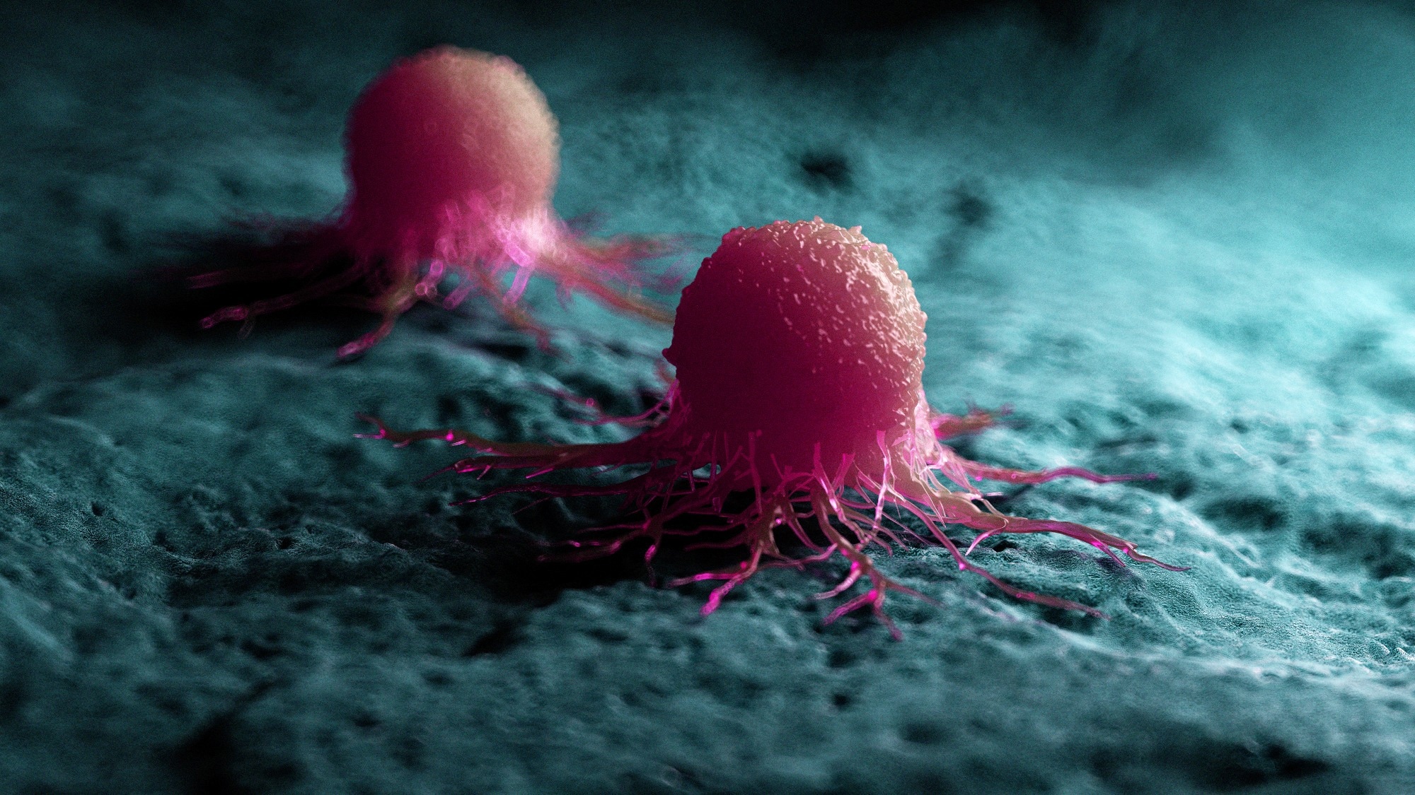
In a recent study published in Developmental Dynamics, a group of researchers used a small interfering Ribonucleic Acid (siRNA) screen coupled with a deep attention network (DAN) to identify and rank genes that control neural crest (NC) functions, such as collective migration in metastatic melanoma.
 Image credit: 3dMediSphere/Shutterstock.com
Each year, the ability of melanoma cells to leave primary tumors and invade surrounding tissues contributes to more than 55,000 global cancer deaths. This collective invasion hinges on leader-follower dynamics, extracellular matrix (ECM) remodeling, and tightly regulated guidance cues. However, deciphering which genes drive these behaviours remains a significant challenge.
High-content imaging and machine learning now make it possible to watch, measure, and model crowd behavior at single-cell resolution. The study also innovated technically using a rubber-stopper plug assay rather than scratch wounds, preserving uniformity and cell integrity for precise measurement of invasion dynamics. Mapping the key regulators could inform therapies that control metastasis and repair NC-related birth defects. Further research should validate these leads in diverse in vivo contexts.
About The Study
Investigators cultured human c8161 metastatic melanoma cells (an aggressive neural-crest-derived line) and HT1080 human fibrosarcoma cells as a mesodermal control. Triplicate wells in collagen-coated, plug-blocked 96-well plates received transfection mixes containing pooled siRNA against 45 candidate invasion genes or non-targeting controls. After 48 hours, plugs were removed to create uniform circular voids. Live confocal imaging captured nuclear fluorescence every 30 minutes for 24 hours at 37°C with 5% carbon dioxide. Custom ImageJ macros quantified cell counts and free-space closure, while TrackMate extracted >10,000 trajectories per well.
Statistical scripts computed fold-change z-scores for wound area and proliferation; Welch’s t-tests flagged significant hits. A DAN was then trained on trajectory data to infer neighbor-influence maps, highlighting spatial regions that most affected a focal cell’s next turn. Secondary assays added recombinant proteins like bone morphogenetic protein 4 (BMP4), endothelial cell-specific molecule 1 (ESM1), fibronectin 1 (FN1), interferon-gamma-induced protein 30 (IFI30), vascular endothelial growth factor C (VEGFC), among others, at 50 ng/mL to test migration rescue or stimulation. Finally, green fluorescent protein-labeled c8161 cells, with or without BMP4 knockdown, were transplanted into stage-12 chick hindbrains to assess in vivo invasion over 48 hours.
Study Results
Silencing 14 of the 45 candidate genes markedly delayed melanoma wound closure while leaving proliferation intact, spotlighting cell motility rather than growth as the rate-limiting step. The top six (BMP4, integrin beta 1 (ITGB1), IFI30, potassium voltage-gated channel subfamily E member 3 (KCNE3), protein kinase C theta (PRKCQ), and RAS guanyl releasing protein 1 (RASGRP1)) cut open-space invasion by 25-45% versus controls (p < 0.01).
Four of these genes, BMP4, ITGB1, KNE3, and RASGRP1, also reduced migration in HT1080 fibrosarcoma cells, but only these four overlapped between cell types. Leader-follower analysis revealed that BMP4, ITGB1, KCNE3, and RASGRP1 each trimmed edge-cell speed by roughly one-third; uniquely, BMP4 also halved directional persistence, causing leaders to meander and decouple from followers.
DAN heatmaps supported these findings, showing that control c8161 cells had the broadest range of neighbour awareness. BMP4 knockdown collapsed this field into a bifocal, front-rear pattern, while RASGRP1 knockdown broadened attention laterally, hinting at compensatory neighbor sensing. In contrast, ITGB1 or KCNE3 loss left the canonical pattern intact, suggesting downstream mechanical, not communicative, deficits. Unexpectedly, silencing disabled homolog 2 (DAB2), carbonic anhydrase 2 (CA2), FN1, or beta-catenin (CTNNB1) radically reshaped attention maps yet did not hamper closure, revealing hidden route plasticity in collective migration mechanisms.
Protein add-back assays confirmed secreted ligands as motility cues. BMP4, ESM1, FN1, IFI30, and VEGFC each accelerated closure by 20-35% in melanoma but not fibrosarcoma cultures. Notably, SERPINI1 significantly increased migration only in HT1080 cells, not c8161.
Recombinant BMP4 nearly doubled the migration of BMP4-depleted cells, proving on-target rescue. Conversely, the BMP antagonist Noggin and BMP4 siRNA reduce in vivo advance of transplanted melanoma clusters through chick neural tissue, slashing median invasion distance from 186 µm to 103 µm when grafted adjacent to rhombomere 4.
Collectively, multivariate ranking distilled the 45-gene list into a high-priority quartet (BMP4, ITGB1, KCNE3, and RASGRP1) that governs leader speed; a duo (BMP4 and RASGRP1) that also rewires neighbor communication; and five secreted proteins that can override intrinsic deficits. These insights span molecular, cellular, and tissue scales, demonstrating that integrative screens can uncover actionable invasion nodes within weeks, not years.
Conclusion
This integrative workflow combined high-throughput siRNA silencing, DAN analytics, and embryo transplantation, pinpointing BMP4, ITGB1, KCNE3, and RASGRP1 as central coordinators of NC-like collective invasion. Their influence lies chiefly in steering and synchronizing leader cells rather than fueling proliferation.
Secreted ligands such as BMP4 and VEGFC amplify motility, raising prospects for therapeutic blockade, whereas exogenous BMP4 rescues migration, hinting at regenerative applications. By translating massive trajectory datasets into intuitive “attention maps,” the study offers a scalable blueprint for decoding and directing complex cell migrations that shape development and drive cancer spread.
In vivo validation was only performed for BMP4, and further investigation is needed to test the functional roles of other candidate genes in embryonic models. The study’s DAN approach also uncovered changes in neighbor influence maps even when migration remained unaffected. Although subtle, this suggests cells may reroute their communication architecture without losing invasive potential. This insight into spatial reprogramming adds a fresh dimension to understanding metastasis and developmental patterning.
Download your PDF copy now!
Journal reference:
Kasemeier-Kulesa JC, Martina Perez S, Baker RE, Kulesa PM. (2025). Identification of neural crest and melanoma cancer cell invasion and migration genes using high-throughput screening and deep attention networks. Developmental Dynamics, 1-18. DOI: 10.1002/dvdy.70059 https://anatomypubs.onlinelibrary.wiley.com/doi/10.1002/dvdy.70059