The ImageXpress™ Pico Automated Cell Imaging System extends beyond traditional digital microscopy, merging high-resolution capture with robust data analytics. Designed for both fluorescence and brightfield applications, this automated platform includes a broad range of pre-built assay templates to minimize setup time and accelerate experimental setup.
Video Credit: Molecular Devices UK Ltd
Equipped with real-time 2D Digital Confocal* deconvolution, automated focusing, live preview modes, multi-wavelength cell scoring, and optional integration with IN Carta® Image Analysis Software, this compact, cost-effective system helps users advance their discoveries.
Get Started Quickly
Streamline laboratory imaging using the icon-based, intuitive CellReporterXpress® Image Acquisition and Analysis Software. Acquire images and extract metrics with only limited training required.
Do More than Cell Counting
Diversify experimental capabilities with more than 25 pre-tuned application modules tailored for apoptosis assays, mitochondrial evaluation, 3D cell models, live-cell time-lapse studies, multi-channel scoring, and neurite outgrowth mapping.
Automate Imaging Affordably
Reduce reliance on centralized core facilities by conducting automated imaging and data quantification right on the benchtop. Customize configurations with digital confocal options, environmental controls, and Z-stack imaging to match one’s precise research needs.
ImageXpress Pico Automated Imaging System Virtual Tour. Video Credit: Molecular Devices UK Ltd
Features
Multiple Imaging Modes
Objectives ranging from 4x to 63x are available for the ImageXpress Pico, supporting colorimetric, brightfield, fluorescence, or real-time 2D Digital Confocal deconvolution capabilities.
Preconfigured Analysis Protocols
More than 25 pre-set evaluation templates span basic cell counts to complex neurite network tracing. Using the click-to-find utility, adjusting measurement parameters is as simple as selecting target cells that match the designated parameters.
Plate-to-Individual Cell View
Results display across several scales, ranging from entire plate layouts down to single-cell details. Extensive analytical display features across well and cellular perspectives enable maximum information extraction from experimental samples.
Z-Stack Acquisition
Capture sharper images for improved feature segmentation via multi-plane Z-stack imaging. Gathering sequential pictures across multiple focal positions preserves greater structural detail than single-plane captures. Workflows can incorporate every focal layer or select specific slices for final projection rendering.
Quickly and Easily Identify Regions of Interest
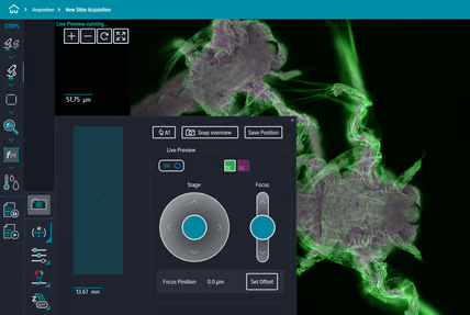
Live Preview streamlines target region detection, enabling smooth sample navigation and manual focus tuning with a virtual joystick, thereby reducing setup time.
Environmental Control
Multi-day, time-series, and live-cell studies run smoothly with integrated environmental hardware that manages humidity, CO2, and O2 levels. Engineered to eliminate focus drift, the interface delivers continuous environmental monitoring to maintain optimal assay conditions.
Different Configurations of the ImageXpress™ Pico
Standard Configuration
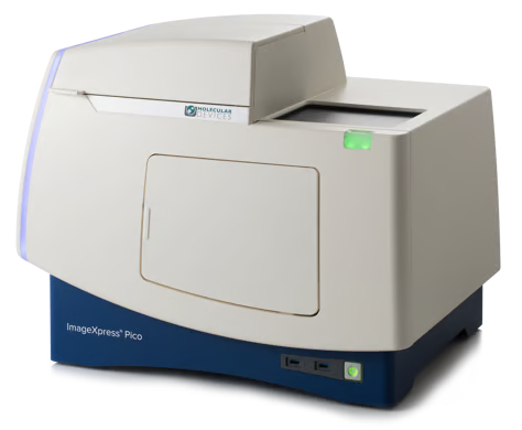
Image Credit: Molecular Devices UK Ltd
Objectives: Leica HC PL Fluotar 4× (NA 0.13) and 10× (NA 0.32); transmitted light capability
Filter cubes: FITC filter assembly designated for fluorescent imaging
Light source: Solid-state illumination system paired with optics, enabling an expanded field of view
Labware: Microtiter plate adapter (six- to 384-well) and four-slide adapter
Software: Includes a CellReporterXpress Software license, supporting five concurrent user access points from desktop or laptop computers, alongside up to two laptop connections per desktop session. This licensing grants access to a maximum of 10 CPU cores reserved for distributed image processing.
Analysis: The standard algorithms for image analysis have 17 analysis modules, including cell counting, cell scoring, apoptosis, and various other analysis modules.
Service: The system includes a one-year MDCare PhD Technical Support Plan and a one-year warranty covering parts and labor.
ImageXpress Pico with Environmental Control
Objectives: Leica HC PL Fluotar 4× (NA 0.13) and 10× (NA 0.32); transmitted light capability.
Filter cubes: FITC filter cube for fluorescence imaging
Light source: Solid-state illumination paired with optimized optics enables an expanded field of view
Environmental Control: Sustains incubation temperatures up to 40 °C, regulates CO2 levels between the ambient atmosphere and 15%, controls O2 levels ranging from 1% to 15% alongside ambient O2, and manages humidity to decrease evaporation.
Labware: Microtiter plate adapter (six to 384-well) and four-slide adapter.
Software: The system is bundled with CellReporterXpress Software licensing, accommodating five concurrent user connections across laptops or desktops, with up to two tablet sessions per active desktop login. It supports parallel image processing across 10 processor cores.
Analysis: Default image evaluation routines encompass 17 specialized processing tools, including cell counting, cell scoring, apoptosis, and other analysis modules
Service: System includes a one-year MDCare PhD Technical Support Plan and a one-year warranty covering parts and labor
Additional Options
Objectives: Leica HC PL FLUOTAR 20× (NA 0.40), 40× (NA 0.60), 63× (NA 0.70)
Filter Cubes: CFP, DAPI, Texas Red, TRITC, Cy5
CellReporterXpress Analysis: Advanced Analysis Module, Z-Stacking, Multi-wavelength Cell Scoring, Translocation Analysis Algorithm, Neurite Tracing Algorithm, Granularity Analysis Algorithm
Metaxpress Analysis:
- MX Basic Software Bundle
- MDCStore Xchange Service
- Translocation
- Angiogenesis
- Neurite Outgrowth
- Cell Scoring
- Count Nuclei
- Granularity
- Live/Dead
- Transfluor
- Cell Health
- Multiwavelength Cell Scoring
- Cell Cycle
- Monopole Detection
- Nuclear Translocation HT
- Cell Proliferation HT
- Micronuclei
- Transfluor HT
- Multiwavelength Translocation
- Mitotic Index
CellReporterXpress Software for ImageXpress Pico
Easy-to-Learn Software Optimized for Automated Digital Microscopy
An intuitive, accessible user interface simplifies quantitative data extraction from automated microscopy images, featuring real-time 2D Digital Confocal deconvolution, automated focusing, and Live Preview for precise region-of-interest selection.
Built to support distributed processing, this software expands analytical throughput, making it an ideal solution for scaling high-content imaging across slides or microplate formats. A linear, icon-based workflow paired with an array of built-in assay protocols ensures a smooth, streamlined user experience.
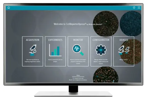
Image Credit: Molecular Devices UK Ltd
IN Carta Image Analysis Software
Modern Machine Learning, High-Content Image Analysis Software
Robust analytics paired with an intuitive interface streamline picture quantification and phenotypic profiling workflows. Advanced capabilities supply essential tools for evaluating 2D, 3D, and 4D datasets at scale, yielding rapid insights without requiring tedious pre-processing or post-processing routines.
Elevate the precision of visual analytics using the SINAP deep-learning framework, proving that 'Segmentation Is Not A Problem'. Tap into machine learning capabilities to conduct sophisticated phenotypic classification through an accessible Phenoglyphs module.
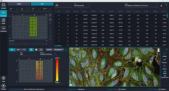
Image Credit: Molecular Devices UK Ltd
Explore the ImageXpress Pico Automated Cell Imaging System in Depth: The System's Front
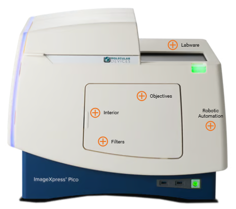
Image Credit: Molecular Devices UK Ltd
Labware
Flexibility to Expand
The ImageXpress™ Pico system supports a variety of laboratory equipment, such as microplates that range from six to 384 wells, both single and multiple slide holders, 35 mm culture dish holders, among others.
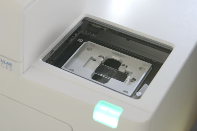
Single Slides. Image Credit: Molecular Devices UK Ltd
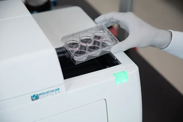
Six-well Cell Culture Plates. Image Credit: Molecular Devices UK Ltd
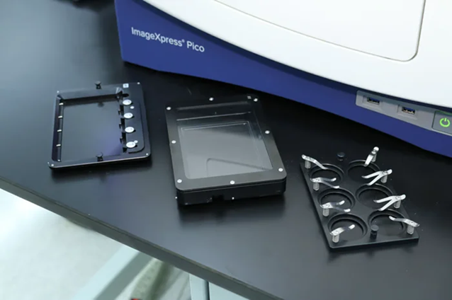
Accommodates a wide variety of labware including a four-slide holder, environmental control cassette, and 35 mm culture dish holders. Image Credit: Molecular Devices UK Ltd
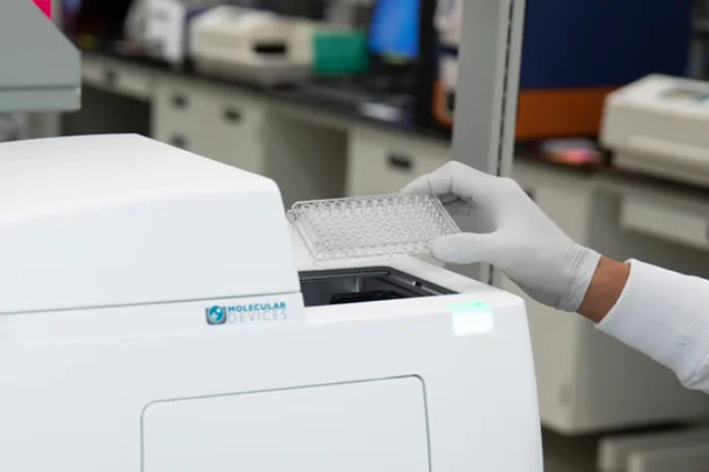
96-well Microplates. Image Credit: Molecular Devices UK Ltd
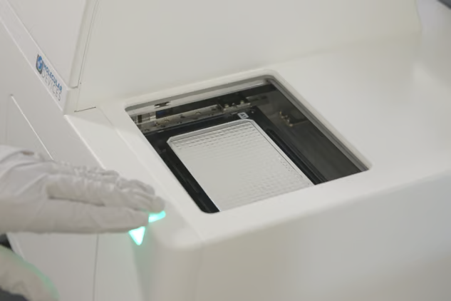
384-well Microplates. Image Credit: Molecular Devices UK Ltd
Objectives
ImageXpress™ Pico incorporates user-exchangeable Leica Microsystems HC PL Fluotar air objectives with extended working distances on an automated turret with six positions.
4× NA 0.13
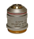
4× NA 0.13. Image Credit: Molecular Devices UK Ltd
4× Leica PL Fluotar Objective (NA 0.13)
Suitable for slides, low-well number plates (six- and 12-well plates), and whole-well imaging.
PLF Fluotars are powerful universal objectives with outstanding chromatic correlation for at least three wavelengths, which makes them suitable for fluorescence imaging. Field planarity (PL) is computed for a 25 mm field of view. They are made of special glass to enable maximum transmission. This makes them powerful photon collectors in fluorescence microscopy. Image Credit: Molecular Devices UK Ltd
10× NA 0.32
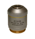
10× NA 0.32. Image Credit: Molecular Devices UK Ltd
10× Leica PL Fluotar Objective NA 0.32 / Free Working Distance: 11.13 mm
The 10× NA 0.32 is suitable for applications involving whole organisms, cell counting (proliferation assessments), live/dead analysis, and apoptosis studies.
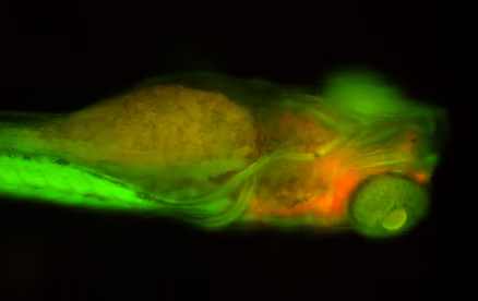
Image Credit: Molecular Devices UK Ltd
20× NA 0.40
20× NA 0.40. Image Credit: Molecular Devices UK Ltd
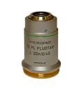
20× Leica PL Fluotar Objective NA: 0.40 / Free Working Distance: 7.45 mm
The 20× NA 0.40 is ideal for cellular, neuron/neurite tracing, puncta counting, and cell scoring.
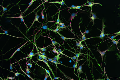
Image Credit: Molecular Devices UK Ltd
40× NA 0.60
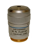
40× NA 0.60. Image Credit: Molecular Devices UK Ltd
40× Leica PL Fluotar Objective NA: 0.60 / Free Working Distance: 3.0 mm Correction Collar
The 40× NA 0.60 is suitable for organelles, puncta counting for small puncta (internalization, endocytosis, phagocytosis), and studies concerning organelles for the purpose of quantifying or examining organelle shape and morphology.
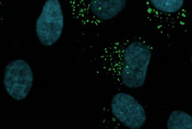
Image Credit: Molecular Devices UK Ltd
63× NA 0.70
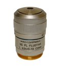
63× NA 0.70. Image Credit: Molecular Devices UK Ltd
63× Leica PL Fluotar Objective NA: 0.70 / Free Working Distance: 2.0 mm Correction Collar
The 63× NA 0.70 is suitable for the assessment of viral titer, imaging of small objects (such as yeast, phytoplankton, and bacteria), as well as visualization of small molecules (including RNA in situ hybridization).
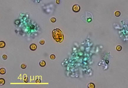
Image Credit: Molecular Devices UK Ltd
Interior
ImageXpress™ Pico employs conveniently accessible filter cubes that can be exchanged by users, along with Leica objectives.
Filters
User-Exchangeable Filter Cubes
The ImageXpress™ Pico Filter Cubes feature a magnetic pin that helps position the filter cube correctly, allowing users to change it with ease.
DAPI Filter Cube
Excitation Range: 350–390 / Emission Range: 419–482
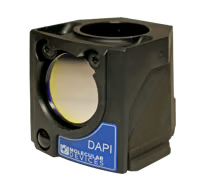
DAPI. Image Credit: Molecular Devices UK Ltd
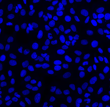
DAPI - 20X U2OS cells with Hoechst-stained nuclei. Image Credit: Molecular Devices UK Ltd
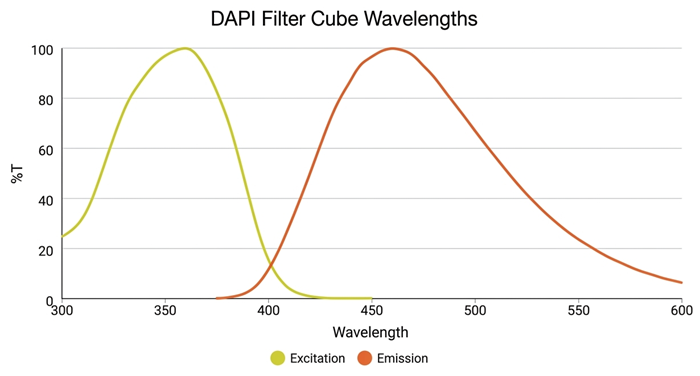
Image Credit: Molecular Devices UK Ltd
CFP Filter Cube
Excitation Range: 350–390 / Emission Range: 419–482
CFP. Image Credit: Molecular Devices UK Ltd
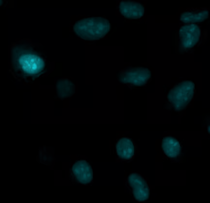
CFP - 40X U2OS cells transduced with bacmam nucleus. Image Credit: Molecular Devices UK Ltd
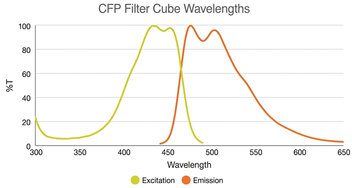
Image Credit: Molecular Devices UK Ltd
FITC Filter Cube
Excitation Range: 445–485 / Emission Range: 509–539
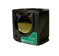
FITC. Image Credit: Molecular Devices UK Ltd
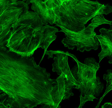
FITC - 40X BPAE cells stained with AlexaFluor 488 phalloidin (actin stain). Image Credit: Molecular Devices UK Ltd
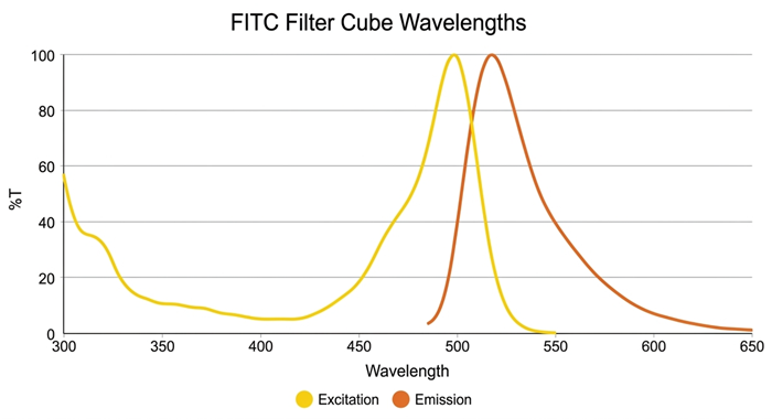
Image Credit: Molecular Devices UK Ltd
TRITC Filter Cube
Excitation Range: 509–552 / Emission Range: 573–613
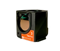
TRITC. Image Credit: Molecular Devices UK Ltd
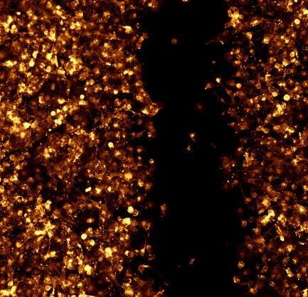
Image Credit: Molecular Devices UK Ltd
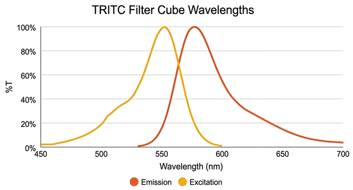
Image Credit: Molecular Devices UK Ltd
Texas Red Filter Cube
Excitation Range: 350–390 / Emission Range: 419–482
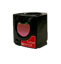
Texas Red. Image Credit: Molecular Devices UK Ltd
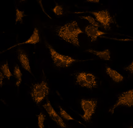
Texas Red - MitoTracker Red stained BPAE cells (mitochondria). Image Credit: Molecular Devices UK Ltd
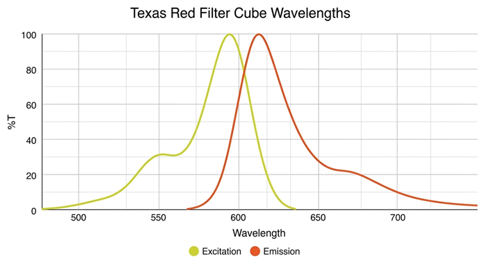
Image Credit: Molecular Devices UK Ltd
Cy5 Filter Cube
Excitation Range: 610–650 / Emission Range: 675–720
Cy5. Image Credit: Molecular Devices UK Ltd
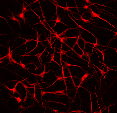
Cy5 - 40X iPSC-derived neurons stained for MAP2B zoomed in. Image Credit: Molecular Devices UK Ltd
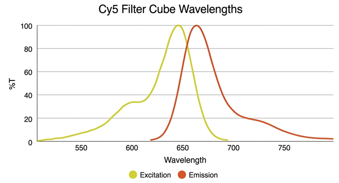
Image Credit: Molecular Devices UK Ltd
Robotic Automation
Customizable to Meet User’s Research Needs
Molecular Devices' ImageXpress™ Pico is specifically designed to work with the S-Lab automated plate handler from PAA. Together, they create a dependable method for unsupervised sample analysis that leads to enhanced productivity, lower expenses, and consistent performance.
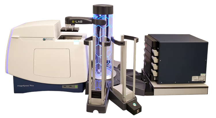
Four-plate incubator shown. Additional configurations available. Image Credit: Molecular Devices UK Ltd
Explore the ImageXpress Pico Automated Cell Imaging System in Depth: The System's Back
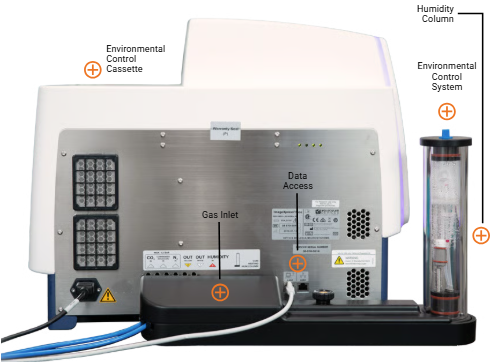
Image Credit: Molecular Devices UK Ltd
Gas Inlet
The device provides users with the capability to conduct live-cell experiments with integrated environmental control.
Environmental Control System
The innovative environmental control cassette chamber, sealed with magnets, regulates gas and humidity levels while minimizing gas consumption as compared to flooding the whole system.
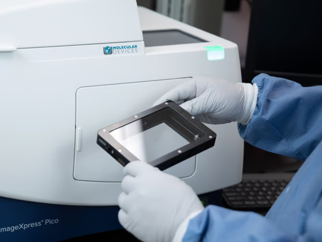
Image Credit: Molecular Devices UK Ltd
Environmental control replicates the conditions found within cells, allowing users to conduct studies over multiple days, observe time-lapse activities, and perform live-cell assays.
Using the CellReporterXpress™ software, the ImageXpress™ Pico Automated Cell Imaging System provides transparency into environmental control parameters throughout the acquisition process, ensuring the system operates correctly.
The environmental management system is embedded directly at the back of the ImageXpress™ Pico, conserving valuable lab bench space. Users can effortlessly introduce water without interrupting current experiments to facilitate studies lasting multiple days.
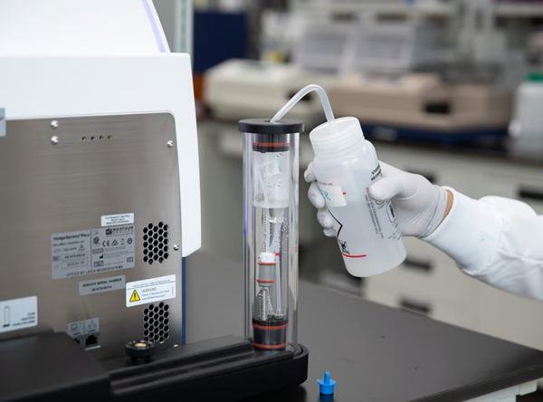
Image Credit: Molecular Devices UK Ltd
Humidity Column
Users can rapidly and effortlessly replenish the built-in humidity column to keep humidity levels at 85% without interrupting the experiment.
Data Access
ImageXpress Pico has integrated CellReporterXpress browser-based software for image acquisition and analysis. The software operates through a web browser and allows users to remotely access their data and manage the ImageXpress™ Pico system from any location, including outside the laboratory.
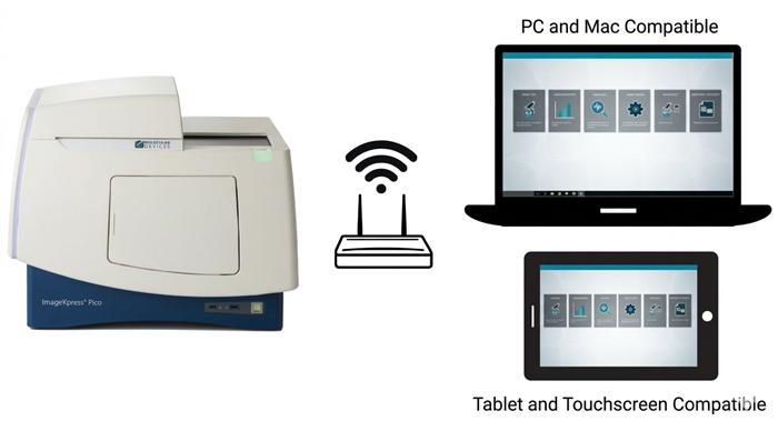
Image Credit: Molecular Devices UK Ltd
Explore CellReporterXpress Software in Depth
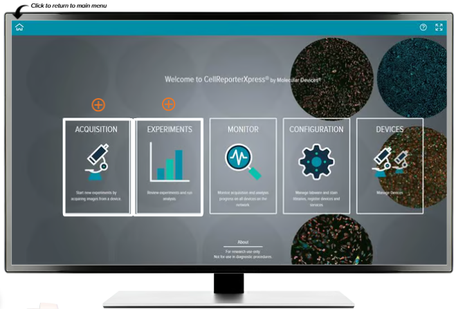
Image Credit: Molecular Devices UK Ltd
Acquisition
The CellReporterXpress has a variety of pre-set protocols–ranging from basic cell counting to complex neurite tracing–designed to capture the clearest images or tailor them to the user’s unique research requirements.
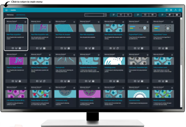
Image Credit: Molecular Devices UK Ltd
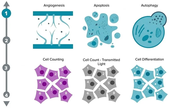
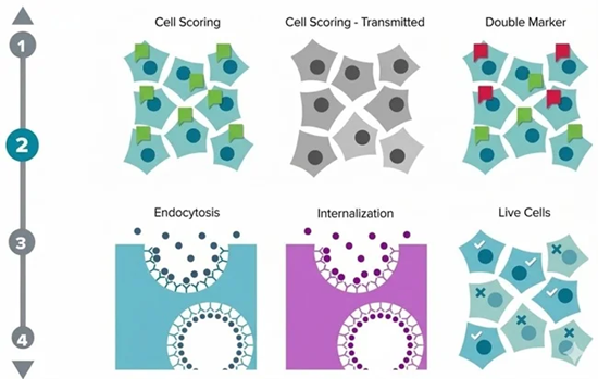
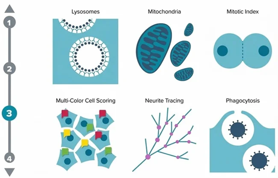
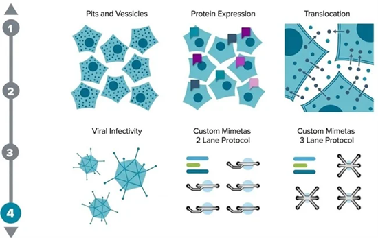
Image Credit: Molecular Devices UK Ltd
Effortlessly navigate configurations to tailor cell image capture to align perfectly with research requirements.
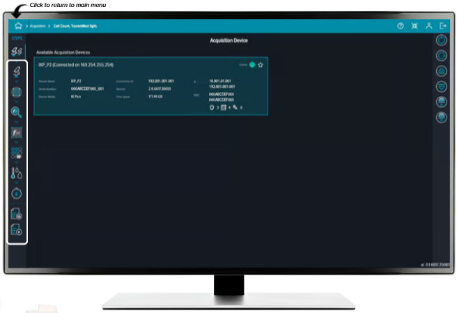
Image Credit: Molecular Devices UK Ltd
Easily Change Plate Format or Accept Defaults
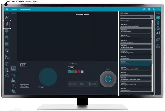
Image Credit: Molecular Devices UK Ltd
The ImageXpress™ Pico system supports a range of labware choices such as slide holders, 35 mm culture dish holders, and additional accessories.
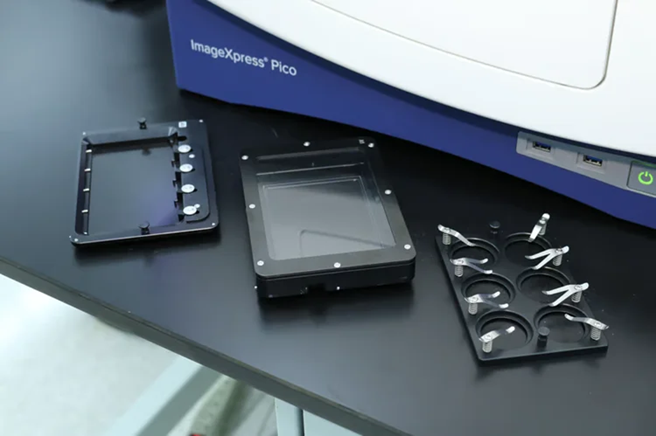
Image Credit: Molecular Devices UK Ltd
Easily Adjust Image Intensity Settings, Including Activating Digital Confocal 2D on-the-fly Image Deconvolution
Digital Confocal Option
Enhance both resolution and assay quality significantly with the Digital Confocal 2D real-time deconvolution. The Digital Confocal feature re-establishes light to its primary source, enabling users to reduce exposure durations and enhance the statistical relevance of their findings.
This system is effortlessly incorporated into Pico's fluorescent imaging workflow, enabling users to obtain fewer z-stacks, which translates to quicker scan rates, along with improved signal-to-noise ratios for more accurate segmentation and evaluation.
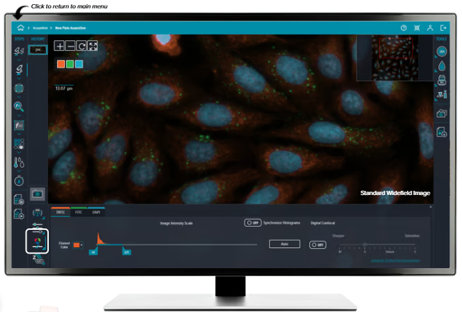
Image Credit: Molecular Devices UK Ltd
Adjust Region Selection Settings Quickly
Live Preview Mode
Facilitate simple recognition of ROIs (regions of interest). Observe the specimen before capture using the virtual joystick to move around it and dynamically adjust the focus. The Live Preview, equipped with a "click-to-center" option, consistently refreshes the image, allowing users to effortlessly explore the specimen to locate the specific field of view they need.
Regardless of whether the user is dealing with 96-well microplates, slides, or 35 mm culture plates, the Live Preview supports users in swiftly and effectively honing in on the key aspects of their research.
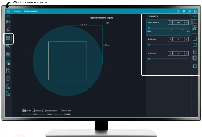
Image Credit: Molecular Devices UK Ltd
Execute real-time analysis by choosing from the list of predefined analysis modules without needing to go back to the main menu.
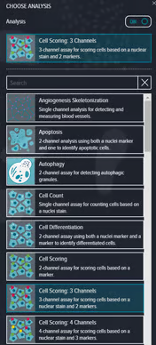
Image Credit: Molecular Devices UK Ltd
Users can analyze the whole acquisition area, or focus on an individual region.
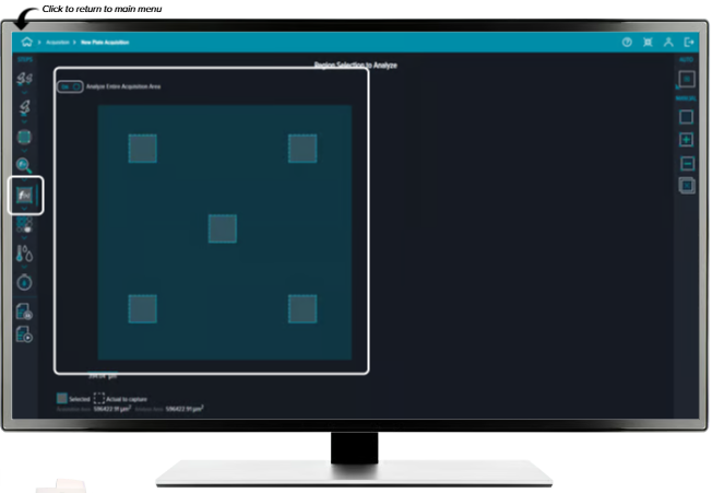
Image Credit: Molecular Devices UK Ltd
Select an entire plate or specific, individual wells.
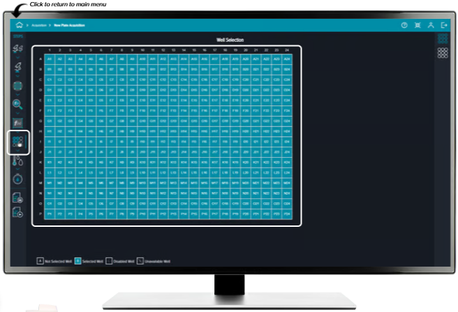
Image Credit: Molecular Devices UK Ltd
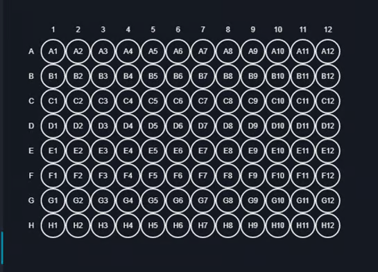
Image Credit: Molecular Devices UK Ltd
Set up a time-lapse series, comprising intermittent time-lapse, to generate movies and dynamic charts.
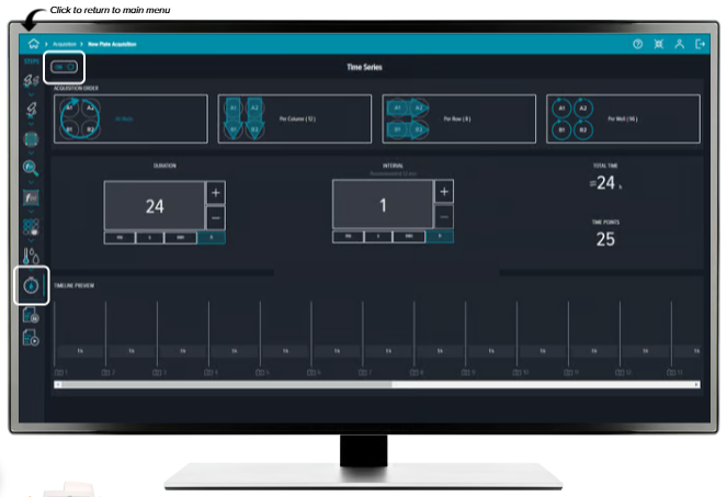
Image Credit: Molecular Devices UK Ltd
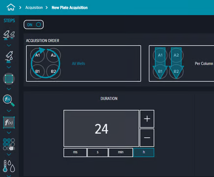
Image Credit: Molecular Devices UK Ltd
Protocol configurations can be stored for effortless future retrieval.
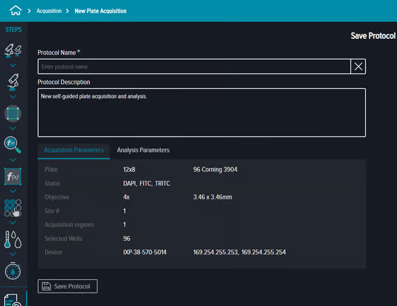
Image Credit: Molecular Devices UK Ltd
After saving, the protocol can be accessed and run from any browser connected to the network.
Experiments
Select from a list, conduct a search, or narrow by specific specifications to locate earlier experiments for review.
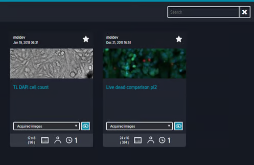
Image Credit: Molecular Devices UK Ltd
Users can examine experimental outcomes from multiple view options (images, plate view, etc.) to effectively meet their requirements.
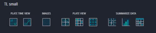
Image Credit: Molecular Devices UK Ltd
Quickly and conveniently switch between data visualization options, such as:
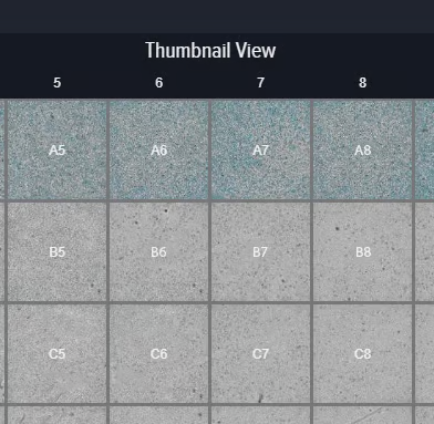
Image Credit: Molecular Devices UK Ltd
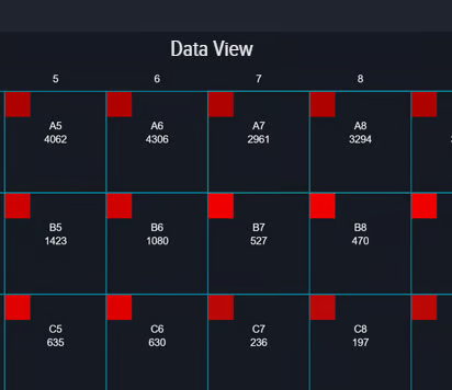
Image Credit: Molecular Devices UK Ltd
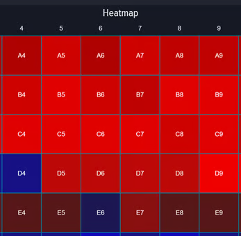
Image Credit: Molecular Devices UK Ltd
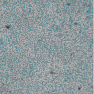
Image Credit: Molecular Devices UK Ltd
The system can automatically produce bar graphs, scatter plots, tables, and more
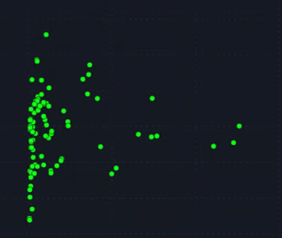
Image Credit: Molecular Devices UK Ltd
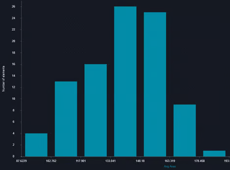
Image Credit: Molecular Devices UK Ltd
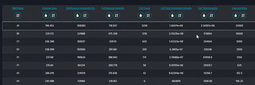
Image Credit: Molecular Devices UK Ltd
Applications of ImageXpress Pico Automated Cell Imaging System
Apoptosis Analysis
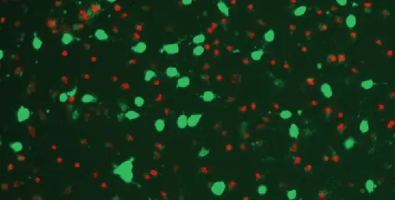
Image Credit: Molecular Devices UK Ltd
Apoptosis is a mechanism of programmed cell death essential for various biological functions, such as embryonic development and routine tissue homeostasis. Impaired apoptotic control plays a central role in diverse pathologies, including malignant conditions.
Specific biochemical cascades induce distinct structural modifications, including cell volume loss, nuclear fragmentation, chromatin compaction, and RNA degradation, eventually culminating in cell death. Apoptosis is triggered through either intrinsic or extrinsic pathways, both of which execute cell degradation by mobilizing caspase enzyme families.
COVID-19 and Infectious Disease Research
Key areas within infectious disease research include host cell engineering, binding kinetics, viral neutralization testing, viral load quantification, and related methods, with a focus on characterizing SARS-CoV-2 to advance COVID-19 management strategies across vaccines, therapeutics, and diagnostic tools.
Cardiomyocytes
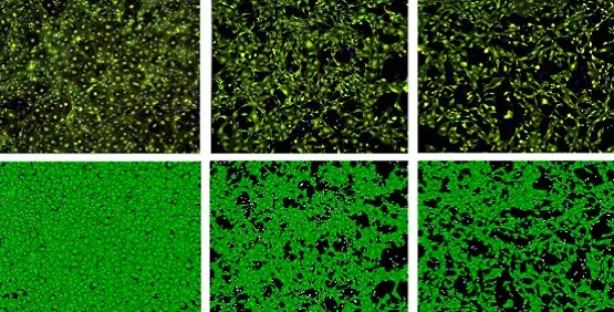
Image Credit: Molecular Devices UK Ltd
Stem-cell-derived cardiomyocytes enable early screening for adverse drug toxicity, preventing investment in therapeutic candidates likely to experience clinical trial failure from cardiotoxic side effects.
Cell Counting
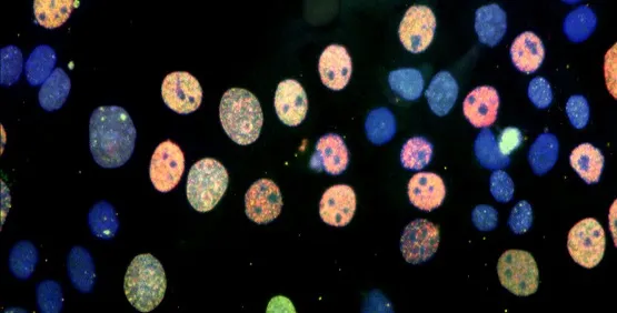
Image Credit: Molecular Devices UK Ltd
Quantifying cells serves as a foundational requirement across diverse biological investigations. Assays measuring chemical toxicity, cellular expansion, and mitotic suppression depend directly on counting cells or population densities in culture wells.
Digital Confocal
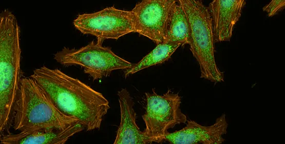
Image Credit: Molecular Devices UK Ltd
Cellular adaptability relies on tightly regulated signaling networks. Extracellular stimuli like hormones or cytokines frequently trigger these pathways, causing cytosolic proteins to migrate into the cell nucleus to modulate target gene expression.
Assays that identify subcellular structures or compartment-specific molecules benefit from image deconvolution, which isolates true fluorescent signals from intense out-of-focus background noise.
The ImageXpress Pico system, equipped with real-time 2D Digital Confocal deconvolution, tracks nucleocytoplasmic protein movement, such as TNF-α-induced NF-κB translocation. Improving image clarity while preserving raw quantitative metrics yields higher statistical power during nuclear translocation analysis.
Disease Modeling
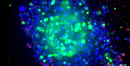
Image Credit: Molecular Devices UK Ltd
Pathological model systems vary in scale and organization, ranging from basic 2D cell cultures to whole-animal models. Whole-organism setups supply in vivo context but entail high costs and often fail to reflect human physiology accurately.
Conversely, standard 2D cultures, despite widespread historical use, cannot reproduce the intricate 3D architecture and intercellular dynamics of intact tissues. Consequently, 3D cell culture systems have established themselves as a compelling alternative for disease modeling.
Drug Discovery and Development
High-content imaging supports pharmaceutical development by monitoring multi-parametric cellular reactions to experimental compounds.
Capturing high-throughput quantitative visual data allows scientists to evaluate structural changes, pathway-specific mechanisms, and cell toxicity throughout preclinical evaluation phases.
Live-Cell Imaging
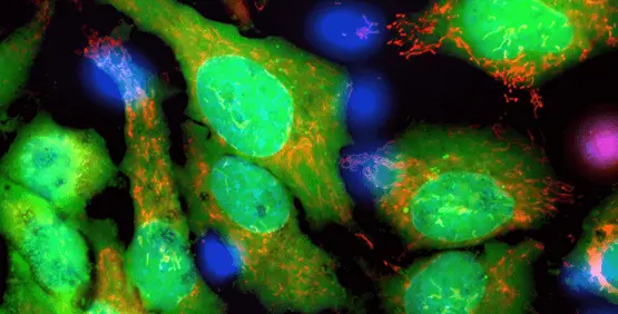
Image Credit: Molecular Devices UK Ltd
Live-cell imaging enables real-time monitoring of physiological activities within viable cells over extended periods under regulated environmental settings.
This methodology allows scientists to track dynamic phenomena, including shifts in cellular structure, motility, or functional responses during an assay. Typical live-cell studies rely on integrated environmental regulation alongside sequential time-lapse image collection.
Neurite Outgrowth/Tracing
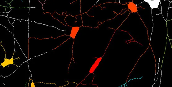
Image Credit: Molecular Devices UK Ltd
Neurite outgrowth quantification evaluates the elongation and branching of neuronal projections throughout differentiation or in response to experimental variables.
High-content microscopy facilitates automated process tracking and parameter extraction, including length, branch density, and structural profile, across cell groups.
Oncology
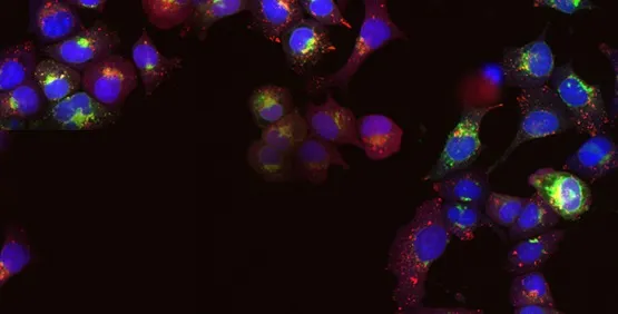
Image Credit: Molecular Devices UK Ltd
Within oncology, high-content imaging examines malignant cell dynamics, structural profiles, and functional changes at single-cell resolution.
This strategy aids the study of mixed cell populations, driving visual quantification of phenotypic diversity, cell cycle progression, or therapy-driven effects in tumor models.
Stem Cell Research
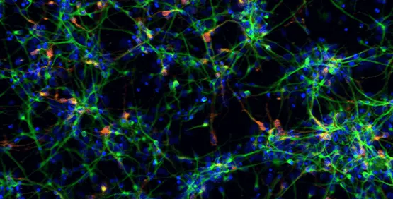
Image Credit: Molecular Devices UK Ltd
Pluripotent stem cells are used for developmental biology investigations or differentiated to generate organ-specific cells, which are then applied in live- or fixed-cell-based assays on slides or in multi-well plates. The ImageXpress system offers value across the entire stem cell researcher's workflow, including differentiation tracking, quality control, and assessing the functionality of particular cell types.
Toxicology
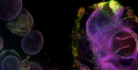
Image Credit: Molecular Devices UK Ltd
Toxicology examines the harmful impacts of natural and synthetic chemical substances on biological systems. It represents an increasing priority in modern research given growing environmental and consumer product exposure to chemical compounds.