The Multi-View Selective-Plane Illumination Microscope (SPIM) from Bruker is not only the fastest light-sheet system on the market, but it also integrates years of Luxendo imaging expertise and invention. Its modular design allows for both live-sample (LS) and cleared-sample (CS) imaging.
MuVi SPIM is the only imaging system that offers high-speed volumetric acquisition of dynamic processes in live specimens as well as cleared sample imaging using any clearing procedure. This is accomplished with a simple and quick objective swap.
Only the Versatile and Customizable MuVi SPIM Provides:
- Concurrent dual-color detection and up to six laser lines 405-to-785 nm
- 360-degree high-resolution artifact-free imaging
- Add-on photomanipulation module
- Complete software support for image fusion, registration, tile stitching, and 3D visualization
MuVi SPIM LS
Optimized for long-term imaging in real-time, with complete environmental control and an optional photomanipulation module.
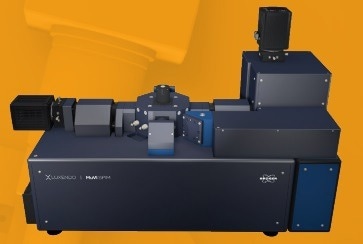
Image Credit: Bruker Nano Surfaces
MuVi SPIM CS
The MuVi SPIM CS preserves optimal image quality, is designed for cleared samples, and supports any type of clearing solution.
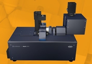
Image Credit: Bruker Nano Surfaces
Why Choose Light-Sheet Microscopy
Light-sheet microscopy is a fluorescence imaging technique that uses a laser light sheet to illuminate only a small fragment of the sample, allowing for inherent optical sectioning. A full-frame camera detects the fluorescence signal of the entire lit 2D plane. This results in distinct imaging benefits:
- Rapid imaging speed
- Low photobleaching and phototoxicity
- High-temporal and 3D-spatial resolution
- Exceptional signal-to-noise ratio
Convenient Multiview Imaging
Absorption and light scattering impair signal and image quality when imaging large samples, inhibiting high-quality imaging. MuVi SPIM is a unique 4-axis concept and comes in a variety of configurations to enable unique properties such as:
- Free rotation axis for optimal sample positioning
- Concurrent illumination and detection from two opposing directions without rotation
- Unrivaled acquisition speed
- Isotropic resolution with a single 90-degree rotation
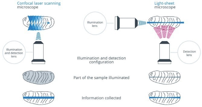
In confocal microscopy the sample is illuminated repeatedly with out-offocus light leading to phototoxic effects and photobleaching. In light-sheet microscopy only the focal region is illuminated, and no unnecessary out-of focus light is generated. Image Credit: Bruker Nano Surfaces
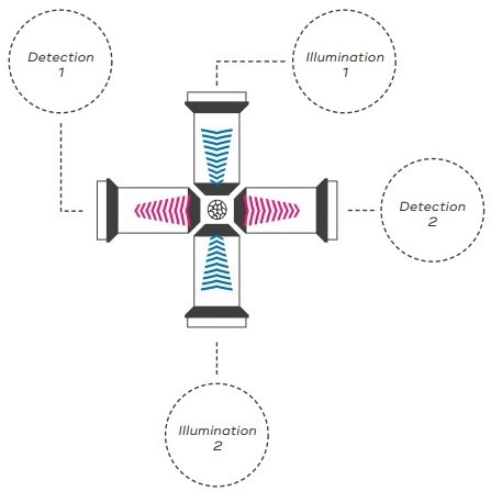
MuVi SPIM 4-axis concept with two opposing detection and illumination objectives. This provides four independent views of the sample, each optimized for one quadrant. Image Credit: Bruker Nano Surfaces
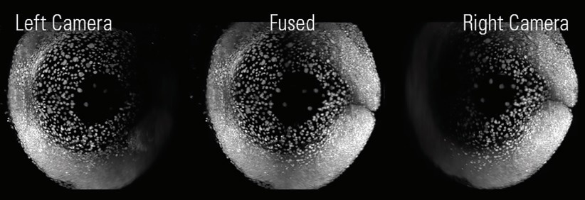
Zebrafish expressing H2A::GFP acquired simultaneously from two opposing directions (left, right). Data fusion of the two views yields a high-quality in-toto image of the sample (middle). Image Credit: Bruker Nano Surfaces
MuVi SPIM LS
Pure Live Imaging
Native and Natural Environment
MuVi SPIM, which includes an environmental control module, can image live specimens for hours or days without affecting biology. This is accomplished by regulating temperature (20–37 °C), gas concentrations (CO2, O2, N2), and humidity, allowing for research in:
- Marine biology
- Oncology
- Developmental biology
- Neurobiology and neurodevelopment
Unparalleled Imaging Speed for In-Toto Imaging
The MuVi SPIM LS can catch a large number of biological events due to its fast-imaging speed. This results in:
- Four combinations of illumination and detection
- 100x to 1000x speedier than confocal microscopy
- Ideal for cellular and subcellular tracking
- Excellent for functional imaging
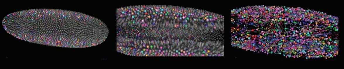
Time series of drosophila development. Cell tracking created with Arivis Vision4D 3D visualization and analysis software. Image Credit: Celia Smits and Stanislav Y. Shvartsman. Department of Molecular Biology at Princeton University, NJ.
Optional Photomanipulation
The ability to regulate, manage, and alter biological processes in space and time is critical for uncovering underlying mechanisms. MuVi SPIM can be enhanced with a photomanipulation (PM) module, which enables advanced studies in photoablation, cauterization, optogenetics, and fluorescence recovery after photobleaching (FRAP) with:
- Diffraction-limited illumination spot
- Beam coupled into detection objective
- Flexible ROI generation (point, straight line, freeform, and square)
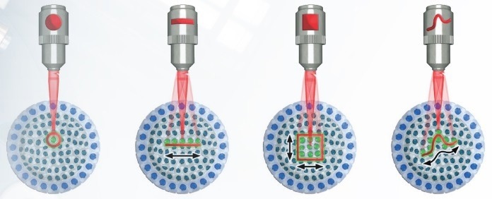
MuVi SPIM provides a variety of excitation schemes and patterns for photomanipulation. CW and pulsed lasers from UV to IR. Image Credit: Bruker Nano Surfaces
MuVi SPIM CS
Comprehensive Cleared-Sample Imaging
Advanced Cleared-Sample Applications
The MuVi SPIM CS has been optimized for cleared samples, allowing for extensive imaging of huge, completely cleared specimens.
MuVi SPIM CS is useful for a wide range of applications and structures, including:
- Brain and central nervous system tissues
- 3D microstructure analysis of tissues
- Organ development
- Tumorigenesis
High Flexibility of Sample Size and Clearing Method
MuVi SPIM CS can image a wide range of cleared samples of varying sizes, from 3D organoids to cleared mouse brains. This is achieved by:
- Unique optical design compatible with all clearing solutions
- 10 × 10 mm area accessible for imaging
- Different configurations of detection lenses to suit the properties of samples
Innovative and Easy Sample Handling
MuVi SPIM CS is an innovative technique for sample mounting that reduces the user's exposure to clearing reagents. This is accomplished by:
- Overhead 3D translation/rotation unit for sample mounting from the top
- Different mounting methods (e.g. hook, plate, pin, cuvette, etc.)
- Effortless maintenance and cleaning of objectives and chamber
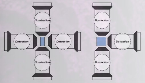
Dual- or single-sided detection combined with dual-sided illumination can be configured according to the experimental needs. Image Credit: Bruker Nano Surfaces
Neurons of a transgenic mouse expressing YFP with the brain being cleared with Clarity. Image Credit: Dr Zhang Dan, Tsinghuan University, China.
Next-Generation Advances
Larger Field-of-View (FOV) and Superior Axial Resolution
MuVi SPIM generates large FOV images with a great 3D spatial resolution by employing a tightly focused Gaussian beam that is swept at high speed along the illumination axis. Integration over time and space results in an extended, uniform, and thin light-sheet with axial resolution over a wide FOV.
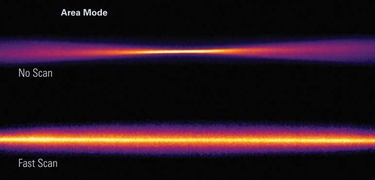
Profile of Gaussian beam generates non-uniform resolution (top). Fast axially scanned beam generates a homogeneous illumination profile. (bottom). Image Credit: Bruker Nano Surfaces
Improved Signal-to-Noise
Light scattering can cause lower contrast and image blur when imaging larger samples or opaque materials. MuVi SPIM has taken an elegant way to improve image contrast by providing:
- Scanned light-sheet
- Line illumination for improved background suppression
- Sophisticated aberration tolerance (even in complex samples)
Effective Elimination of Striping Artifacts
Obstacles such as pigments or cell nuclei can absorb or scatter light while imaging live, cleared, or fixed biological samples, resulting in images with shadows and striping artifacts. The MuVi SPIM uses dual de-striping via pivot scanning to provide a homogenous illumination profile without compromising acquisition speed.
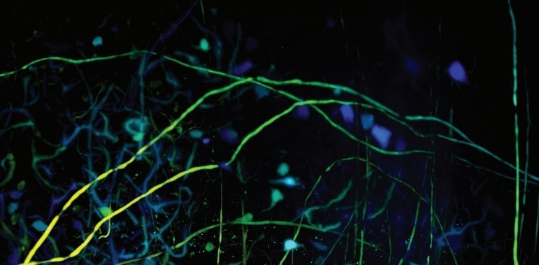
Mouse neurons color-coded by depth, showing high signal-to-noise ratio. Image Credit: Bruker Nano Surfaces
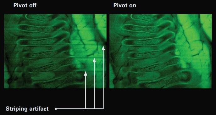
Cleared mouse embryo shows striping artifacts without pivot scanning (left). With pivot scanning, artifacts can be successfully removed (right). Image Credit: Bruker Nano Surfaces
All-in-One Software
Intuitive Design
Bruker’s user-friendly LuxControl software simplifies the setup and performance of multidimensional experiments by providing:
- High reproducibility of experiments: parameters and configurations are saved in the metadata
- All-in-one intuitive user interface: acquisition, viewer, image processor
- Completely scriptable microscope control and processing via an open interface
- Data formats (.tiff, .hdf5, .ims) compatible with common image processing software
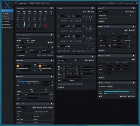
LuxControl image acquisition tab with experimental settings. Image Credit: Bruker Nano Surfaces
3D Data Viewer
LuxControl includes a 3D data viewer that enables scientists to see the complete dataset immediately after acquisition.
- Capability to turn stitching of tiles on or off
- Flexible options to draw and annotate
- Both raw and post-processed images
- A quick viewing of multi-terabyte data sets
Impressive Image Processor
MuVi SPIM records the sample from several angles and is made up of many tiles. The LuxControl software handles image post-processing tasks, such as registration and fusion, which compensate for absorption and scattering and give high-quality data.
Features include:
- Multi-color alignment
- Multi-view image fusion and deconvolution
- Tile stitching of hundreds of tiles for large samples
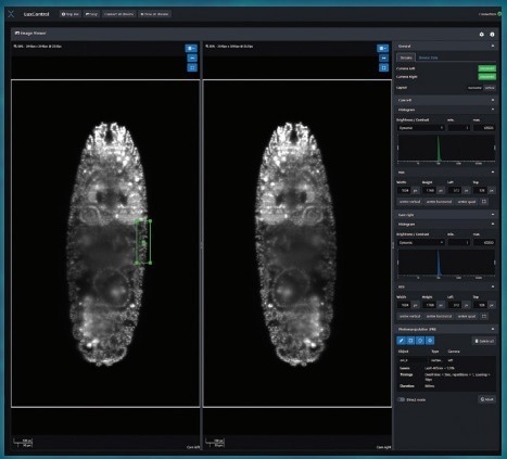
Image viewer and user ROI selection. Image Credit: Bruker Nano Surfaces
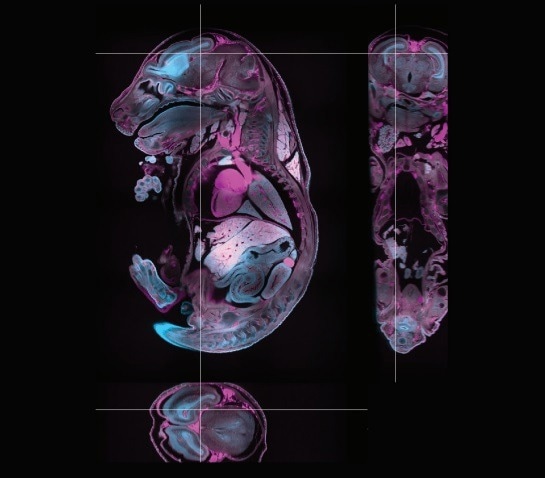
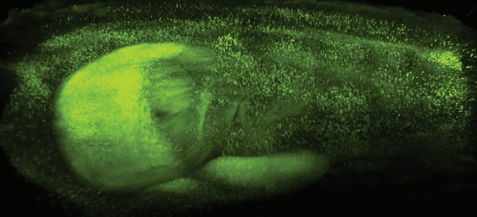
Cleared mouse embryo labeled with methylene blue (cyan) and showing autofluorescence (magenta). Image composed of 12 tiles with 920 planes, processed and stitched using the LuxBundle Image Processor. Image Credit: Montserrat Coll Lladó, European Molecular Biology Laboratory, Barcelona, Spain.
Specifications
Source: Bruker Nano Surfaces
MuVi LS
| Detection Objective |
Effective Magnification |
Field of View µm* |
Pixel Size nm |
| 20x 1.0NA |
11.1x |
1198 |
585 |
| |
22.2x |
599 |
293 |
| |
33.3x |
399 |
195 |
| |
44.4x |
299 |
146 |
| 16x 0.8NA |
8x |
1664 |
812 |
| |
16x |
832 |
406 |
| |
24x |
554 |
271 |
| |
32x |
416 |
203 |
* Calculated based on chip size.
MuVi CS
| Detection Objective |
Effective Magnification |
Field of View µm* |
Pixel Size nm |
| 20x 1.0NA |
10x |
1331 |
650 |
| |
20x |
665 |
325 |
| |
30x |
444 |
217 |
| |
40x |
333 |
163 |
| 10x 0.5NA |
5x |
2662 |
1300 |
| |
10x |
1331 |
650 |
| |
15x |
887 |
433 |
| |
20x |
666 |
325 |
| 4x 0.28NA |
2.2x |
5990 |
2925 |
| |
4.4x |
2995 |
1463 |
| |
6.7x |
1997 |
975 |
| |
8.9x |
1497 |
731 |
* Calculated based on chip size.