There are three models in the HT7800 series: the HT7800, HT7820, and HT7830. All three microscopes usher in a new age of transmission electron microscopy, combining Hitachi's 75+ years of expertise with cutting-edge digital technology. This series, intended for both experienced researchers and novices, provides high-resolution imaging with a user-friendly interface and ergonomic controls that improve efficiency and comfort.
The HT7800 series is designed for life sciences, material sciences, and industrial applications. It offers diverse imaging modes, high-speed digital processing, and seamless automation to ensure precise and repeatable results.
Users can also use the microscope under regular room lighting conditions, eliminating the requirement for a darkroom and improving use and comfort.
- Dual-Mode objective lens: Users can switch between high-contrast and high-resolution modes with a single click, providing unparalleled versatility
- User-friendly interface: Under ambient room lighting conditions, a modern, user-friendly GUI enables smooth operation
- Improved digital imaging system: Low-dose capabilities enhance sample integrity
- Expandable features: MirrorCLEM, EDX, STEM, and more specialized attachments can be integrated for various research requirements
Features and Benefits
Dual-Mode Objective Lens for Maximum Versatility
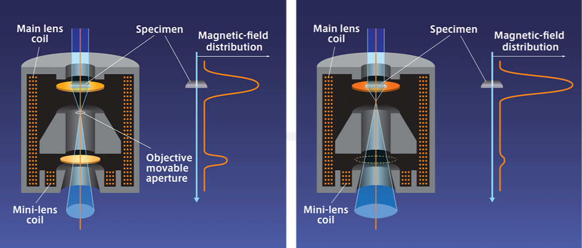
Image Credit: Hitachi High-Tech Europe
- Users can effortlessly transition between high-contrast and high-resolution modes in one microscope
- This series improves workflow efficiency and cost-effectiveness by doing away with the need for several systems
Intuitive Operation and User-Friendly Interface
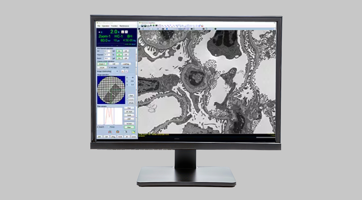
Image Credit: Hitachi High-Tech Europe
- With its simplified digital graphical user interface, the HT7800 series can be used easily by both inexperienced and seasoned users
- With the built-in CMOS screen camera, users can work in a well-lit environment, which will improve ergonomics and lessen fatigue
The HT7800’s Digital Imaging System
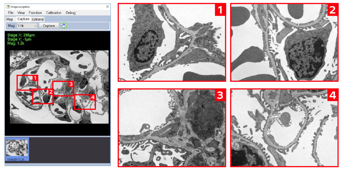
Image Credit: Hitachi High-Tech Europe
- CMOS high-speed camera for better imaging
-
Whole View Function allows rapid, automated image capture across wide sample areas.
Automation and Navigation
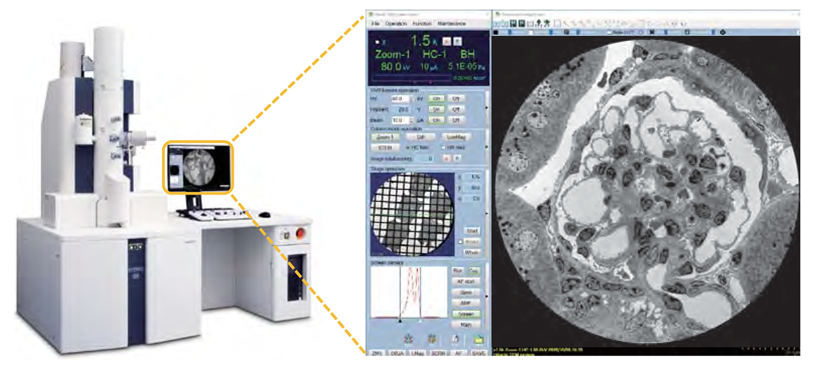
Image Credit: Hitachi High-Tech Europe
Auto Multiple Frame (AMF) imaging: Able to produce high-resolution panoramic imaging by rapidly stitching together numerous images
Advanced image mapping and navigation: Enables finding and examination of regions of interest quickly
3D electron tomography: Produces precise 3D reconstructions by capturing tilted images
Expandable and Customizable for Advanced Research Needs
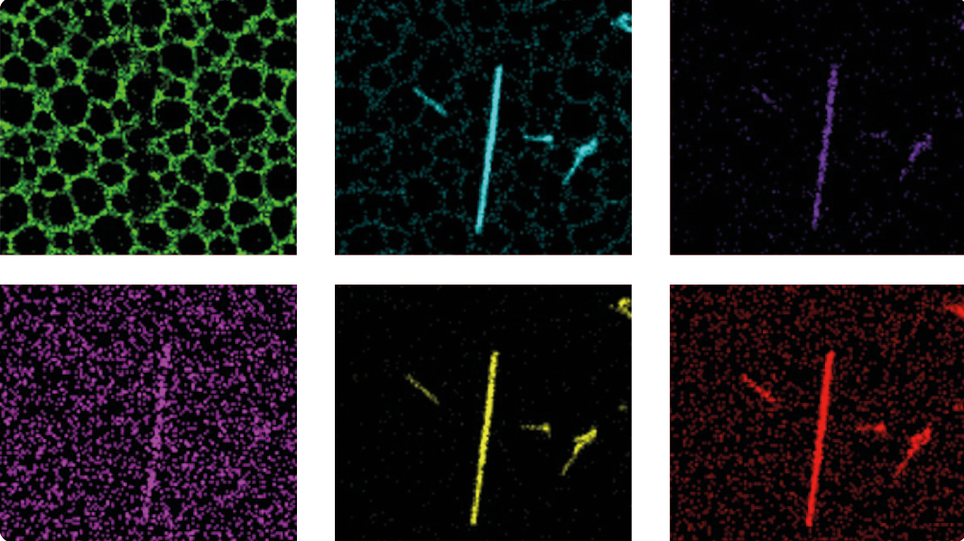
Image Credit: Hitachi High-Tech Europe
STEM & EDX: These methods facilitate material characterization and elemental analysis.
MirrorCLEM technology: Ability to correlate TEM imaging and fluorescence microscopy to provide thorough research findings.
Specifications
Source: Hitachi High-Tech Europe
| |
HT7800 |
HT7820 |
HT7830 |
| Electron gun |
W (standard), LaB6 |
LaB6 (standard), W |
LaB6 (standard), W |
| Accelerating voltage |
20-120 kV (100 V/step variable) |
20-120 kV (100 V/step variable) |
20-120 kV (100 V/step variable) |
| Resolution (Lattice) |
0.20 nm (Off-axis, 100 kV) |
0.14 nm (Off-axis, 120 kV) |
0.14 nm (Off-axis, 120 kV)
0.19 nm (On-axis, 120 kV) |
| Maximum magnification |
x600,000 |
x800,000 |
x1,000,000 |
| Stage maximum tilt angle |
±70° |
±30° |
±10° |
Standard
features |
Auto focus, Microtrace, Autodrive, Live FFT display, Measurement function, Low dose, API (auto pre-irradiation),
Image navigation function, Column with mild baking function, Whole view function, Drift correction function, etc. |
Application:
Life Sciences & Biomedical Research
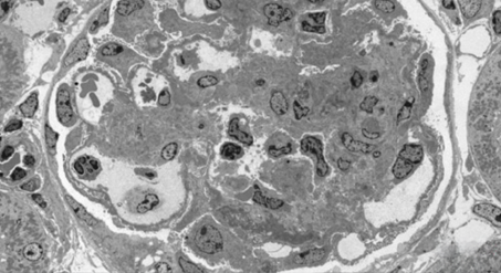
Mouse Kidney (stained), accelerating voltage 80 kV, magnification x300. Image Credit: Hitachi High-Tech Europe
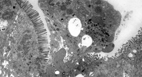
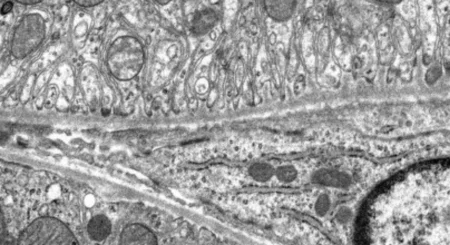
Rat jejunum (unstained), accelerating voltage 80 kV, magnification x2,000. Image Credit: Hitachi High-Tech Europe
- The images above compare an unstained segment using the HT7800 series High Contrast lens with a conventionally stained section
- High contrast imaging for biological materials
- Low-dose mode for sensitive biological specimens and cryo-TEM
Material Science and Nanotechnology
Au/TiO2 catalyst. Bright-field TEM image, accelerating voltage 120 kV. Image Credit: Hitachi High-Tech Europe
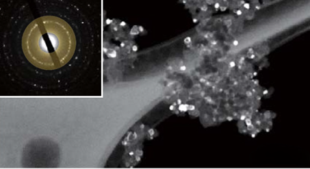
Au/TiO2 catalyst. Hollow-cone dark-field TEM image, accelerating voltage 120 kV. Image Credit: Hitachi High-Tech Europe
- The image above shows hollow-cone dark-field observation using an Au/TiO2 catalyst on the HT7820. The electron beam diffraction region used for the hollow-cone dark-field measurements is highlighted in yellow in the selected-area electron diffraction pattern. TiO2 diffraction waves may be seen clearly in the hollow-cone dark-field photos
- High-resolution imaging of innovative materials, polymers, and nanoparticles
- STEM & EDX compatibility for compositional analysis
Particle/Polymer
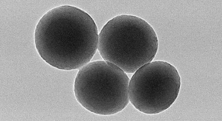
Mesoporous silica powder, accelerating voltage 120 kV, magnification x70,000. Image Credit: Hitachi High-Tech Europe
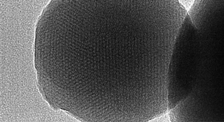
Mesoporous silica powder, accelerating voltage 120 kV, magnification x200,000. Image Credit: Hitachi High-Tech Europe
- The images above demonstrate the TEM observation results of mesoporous silica particles, which are likely to be used in drug delivery systems
- High-resolution performance and digitization enable users to view the sequence of multiple nanometer pores immediately.