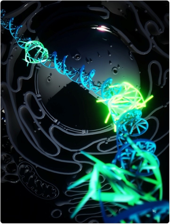
Quadruple helix DNA (green) forming. Image Credit: Ella Maru Studio.
DNA generally forms the traditional double helix shape that was identified earlier in 1953—a pair of strands wound around one another. While many other structures have been created in test tubes, this does not essentially imply that they develop inside the living cells.
Termed DNA G-quadruplexes or G4s, quadruple helix structures have earlier been identified in cells. But in this method, either the cells are killed or high concentrations of chemical probes are required to view the formation of G4; hence, their actual presence inside the living cells under usual environmental conditions has not been monitored, to date.
Now, a team of researchers from the University of Cambridge, Imperial College London, and Leeds University has developed a fluorescent marker that can bind to G4s found in living human cells, enabling them to observe the formation of the structure and the type of role it plays in cells, for the first time.
The study was recently published in the Nature Chemistry journal.
Rethinking the biology of DNA
Dr Marco Di Antonio is one of the lead scientists who started the research in the laboratory of Professor Sir Shankar Balasubramanian at the University of Cambridge. Dr Di Antonio presently heads a team of researchers in the Department of Chemistry at Imperial College London.
For the first time, we have been able to prove the quadruple helix DNA exists in our cells as a stable structure created by normal cellular processes. This forces us to rethink the biology of DNA. It is a new area of fundamental biology, and could open up new avenues in diagnosis and therapy of diseases like cancer.”
Dr Marco Di Antonio, Lead Researcher, Department of Chemistry, Imperial College London
Dr Di Antonio continued, “Now we can track G4s in real time in cells we can ask directly what their biological role is. We know it appears to be more prevalent in cancer cells and now we can probe what role it is playing and potentially how to block it, potentially devising new therapies.”
The researchers believe that G4s form in DNA to momentarily keep it open and enable processes, such as transcription, in which the DNA instructions are read followed by protein synthesis. This is a kind of “gene expression” in which a portion of the genetic code in the DNA is stimulated.
It appears that G4s are linked more often to genes that play a role in cancer. They are found in huge numbers inside tumor cells. With the potential to presently image a single G4 at a time, the researchers believe that they can possibly monitor the function of the G4s inside specific genes and the way these express in cancer. This underlying knowledge could lead to novel targets for drugs that disrupt the process.
Natural formation
This advancement to image single G4s came with a rethink of mechanisms that are often used to explore the function of cells. Earlier, the scientists had employed molecules and antibodies that could identify and adhere to the G4s; however, these required extremely high levels of the “probe” molecule.
In other words, the probe molecules could be disturbing the DNAs and actually forcing them to form G4s, rather than identifying them while they naturally form.
Dr Aleks Ponjavic, currently an academic in the Schools of Physics & Astronomy and Food Science and Nutrition at the University of Leeds, mutually headed the study in the laboratory of Professor Sir David Klenerman and devised the technique for observing the novel fluorescent marker with microscopy.
Scientists need special probes to see molecules within living cells, however these probes can sometimes interact with the object we are trying to see. By using single-molecule microscopy, we can observe probes at 1000-fold lower concentrations than previously used.”
Dr Aleks Ponjavic, Academic, Schools of Physics & Astronomy and Food Science and Nutrition, University of Leeds
Aleks Ponjavic continued, “In this case our probe binds to the G4 for just milliseconds without affecting its stability, which allows us to study G4 behaviour in their natural environment without external influence.”
For the latest probe, the researchers employed a highly “bright” fluorescent molecule in negligible proportions, which was made such that it adheres quite easily to the G4s. The insignificant amount meant that the researchers could not expect to image each G4 in a cell, but can rather detect and monitor single G4s, enabling them to figure out their underlying biological function without disturbing their overall stability and prevalence in the cell.
The scientist successfully demonstrated that G4s seem to develop and disintegrate very rapidly, indicating that they form only to carry out a particular function, and that potentially if they lasted long enough, they can turn toxic to normal processes of the cell.
Source:
Journal reference:
Di Antonio, M., et al. (2020) Single-molecule visualization of DNA G-quadruplex formation in live cells. Nature Chemistry. doi.org/10.1038/s41557-020-0506-4.