RIKEN scientists have developed a new, multipurpose probe that is capable of detecting the programmed destruction of faulty mitochondria—the powerhouses of cells—with pinpoint precision.

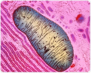
Colored transmission electron micrograph of a single mitochondrion in a human pancreas cell. RIKEN researchers have developed a fluorescent probe that can detect the programmed death of defective mitochondria in the lysosomes. Image Credit: K.R. Porter/ Science Photo Library.
The team used the new probe to demonstrate that impaired mitochondria in neurons, which produce the neurotransmitter dopamine, are not damaged in mice that exhibit a condition mimicking Parkinson’s disease.
Mitochondria are, essentially, organelles that produce most of the chemical energy required by human cells to work. However, during cellular stress, mitochondria can breakdown and generate extremely reactive oxygen radicals, which harm the cells.
Therefore, cells regularly detect and kill dysfunctional mitochondria by assigning them to lysosomes, which serve as a cellular waste-disposal system, by disintegrating unnecessary components.
However, if this selective removal of defective mitochondria—called mitophagy—fails, it can result in numerous diseases. Hence, there is a great deal of interest in tracking cellular mitophagy.
The main reason for developing the fluorescent probes is to identify mitophagy. However, some of these probes can only be utilized in living cells, while others are susceptible to destructive procedures in which lysosomes are not involved.
Atsushi Miyawaki from the RIKEN Center for Brain Science and co-workers have now created a novel fluorescent probe that can be applied to both fixed and living cells and has been particularly highlighted in lysosomes.
The new probe consists of two parts—one part can resist the enzymes in the lysosome, while the other one is damaged by them. Therefore, by tracking the color of the fluorescence of the probe, the team could identify when a mitochondrion had penetrated a lysosome.
The new probe is different from their previous mitophagy probe and is susceptible to acidity and the degrading enzymes found in lysosomes; hence, it functions even in fixed cells in which lysosomes are not acidic anymore.
The researchers employed the new probe to study Parkinson’s disease—a kind of neurodegenerative disorder that causes muscle stiffness, shaking, and progressive movement difficulties.
The team used a mouse model of Parkinson’s disease and identified that dopamine-producing neurons failed to remove the dysfunctional mitochondria; however, other neurons that did not generate dopamine were able to do so.
A dopamine deficiency in the brain can lead to Parkinson’s disease. This, thus, indicates that the inability of dopamine-producing neurons to carry out mitophagy could be a crucial factor in this disorder.
Miyawaki’s research team, in collaboration with scientists from the pharmaceutical company Takeda, detected a compound that can promote the destruction of impaired mitochondria. Compounds like these could help treat Parkinson’s disease in the days to come.
The new probe is a potential tool for improving studies in other diseases.
Since many other neurodegenerative disorders involve mitophagy, our probe can contribute to their study. Furthermore, diseases in other organs involve oxidative stress and hence mitophagy. We’re currently using our probe to look at heart disease.”
Atsushi Miyawaki, Study Corresponding Author, RIKEN
Source:
Journal reference:
Katayama, H., et al. (2020) Visualizing and Modulating Mitophagy for Therapeutic Studies of Neurodegeneration. Cell. doi.org/10.1016/j.cell.2020.04.025.