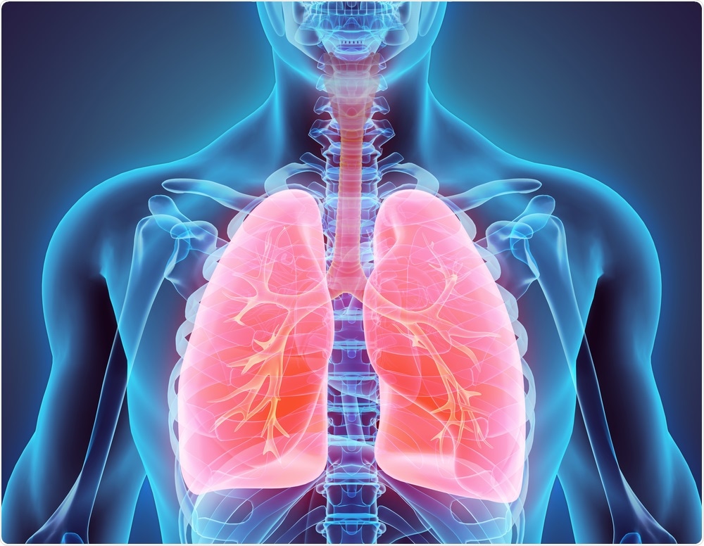
Scientists from Children’s Hospital of Philadelphia (CHOP) and the Perelman School of Medicine from the University of Pennsylvania have discovered a new cellular pathway that can be targeted with a naturally occurring medication to activate the regeneration of lung tissue, which is crucial to recover from multiple lung injuries.

Image Credit: MDGRPHCS/Shutterstock.com
Published recently in the Nature Cell Biology journal, the findings could result in better treatments for lung disease patients, including acute respiratory distress syndrome (ARDS) caused by COVID-19.
Using cutting-edge technology, including genome-wide and single-cell analyses, we have identified a specific cellular pathway involved in lung tissue regeneration and found a drug that enhances this process. These findings provide identification of precision targets and thus allow for rational development of therapeutic interventions for lung disease caused by COVID-19 and other illnesses.”
G. Scott Worthen, MD, Study Senior Author and Physician-Scientist, Division of Neonatology, Children’s Hospital of Philadelphia
Dr Worthen is also a member of the Penn-CHOP Lung Biology Institute.
Conditions like influenza, pneumonia, and ARDS—one of the familiar complications of COVID-19 disease—can affect the lining of the air sacs in the lungs, called the alveolar epithelium, which inhibits oxygen from moving from the lungs to the bloodstream and can result in death.
COVID-19 patients who develop ARDS become seriously ill, and so far, no medications have been specifically developed to treat ARDS in COVID-19 patients. Interpreting which genetic pathways and targets are involved in regenerating epithelial tissue is crucial in developing effective treatments for ARDS and analogous conditions.
An earlier study has demonstrated that type II alveolar pneumocytes (AT2) are major cells involved in lung repair, both via self-renewal and transdifferentiation into type I alveolar pneumocytes (AT1), which promote gas exchange between the lung air sacs and neighboring capillaries.
Yet before this research, scientists were unaware of the type of gene accessibility changes that occurred in AT2 cells after disease-related injury to support repair, and they were also clueless about the regenerating AT2 cells and the way they influence the interactions with neighboring mesenchymal cells, which are also significant in tissue repair.
Through using genome-wide analyses, the researchers evaluated changes in the AT2 cells following lung injury, which opens up the chromosomes inside the cells and makes certain genes available to the cellular machinery.
Subsequently, the team employed single-cell analysis of mesenchymal cells and AT2 cells to better interpret the way these two cell types communicate during injury and the type of cell signaling pathways that are involved.
Both methods converged on a single route, wherein a transcription factor called STAT3 enhanced the expression of brain-derived neurotrophic factor (BDNF), which consequently increased the regeneration of lung tissue.
Upon further analysis of this pathway, the team detected a naturally-occurring compound called 7,8-Dihydroflavone (7,8-DHF), which targeted a receptor in the pathway, activating and expediting lung tissue repair in numerous mouse models of lung injury.
We believe these findings could lead to the development of a new therapeutic that could help patients recovering from COVID-19 and similar diseases. Based on the results of this study, we think 7; 8-DHF is an excellent candidate for entering clinical trials for patients with lung diseases.”
Andrew J. Paris, MD, Study First Author, Instructor of Medicine, and Pulmonary Specialist, Perelman School of Medicine, University of Pennsylvania
Source:
Journal reference:
Paris, A. J., et al. (2020) STAT3–BDNF–TrkB signalling promotes alveolar epithelial regeneration after lung injury. Nature Cell Biology. doi.org/10.1038/s41556-020-0569-x.