Reviewed by Lexie CornerMay 15 2025
Tissues are composed of a diverse array of cell types. As a result, studying their biological functions and investigating disease mechanisms can be challenging. A multi-institutional team led by Osaka University has developed and demonstrated a proof-of-concept technique that visualizes the distribution of components within individual cells, paving the way for a deeper understanding of disease in complex biological samples.
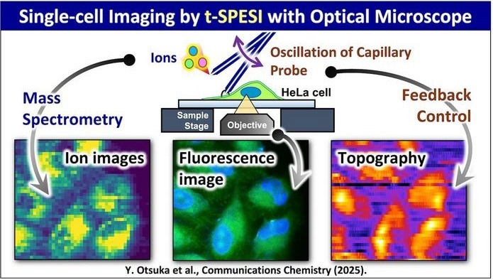
 Schematic diagram of multimodal single-cell MSI using tapping-mode scanning probe electrospray ionization. Image Credit: Yoichi Otsuka
Tapping-mode scanning probe electrospray ionization (t-SPESI) is a technique used to analyze the spatial distribution of molecules within a sample. It works by collecting multiple micro-samples from different parts of a cell and transporting them for analysis via mass spectrometry, which identifies the specific chemical components present in each location.
We have developed a new t-SPESI unit that allows us to visualize the microscopy sample in multiple modes. We can also directly observe the sampling process as the micro-samples are taken for mass spectrometry analysis.
Yoichi Otsuka, Study Lead Author and Associate Professor, The University of Osaka
The research team enhanced their previously developed t-SPESI system by enabling the analytical unit to be mounted above an inverted fluorescence microscope. This modification allows researchers to observe both the sampling process and the sample in real time. The sample can be scanned in various ways, including detecting fluorescently labeled target molecules, mapping features on the cell surface, and imaging the spatial distribution of the cell’s chemical components.
This technique is particularly useful for visualizing the distribution of intracellular lipids - fatty molecules involved in key metabolic processes. Disruptions in lipid distribution and function have been associated with a range of diseases.
When we applied our technology to model cells, we were able to observe the lipids within each individual cell using mass spectrometry imaging, directly visualize the cell by fluorescence microscopy, and also determine the surface shape of the cell.
Michisato Toyoda, Study Senior Author, The University of Osaka
The researchers were also able to distinguish between different types of cells based on their unique molecular compositions.
“This allows an understanding of the multidimensional molecular information of individual cells within a sample of diseased tissue,” added Otsuka.
This emerging technology offers a powerful new approach to studying the mechanisms underlying disease development in complex biological samples. By enabling detailed analysis of the diverse cell types and their interactions within tissues, it holds promise for advancing drug discovery and the development of more precise diagnostic tools across a wide range of diseases.
Source:
Journal reference:
Otsuka, Y., et al. (2025) Single-cell mass spectrometry imaging of lipids in HeLa cells via tapping-mode scanning probe electrospray ionization. Communications Chemistry. doi.org/10.1038/s42004-025-01521-2.