 By Pooja Toshniwal PahariaReviewed by Lexie CornerMay 19 2025
A recent study in Nature Communications highlights a key challenge in single-cell proteomics: protein leakage. Focusing on murine tracheal cells, the research team examined how leakage can skew protein quantification and, by extension, the biological insights derived from these studies.
By identifying and removing permeabilized cells, those that have lost proteins due to membrane damage, from analysis, researchers can significantly improve data quality and reliability. Additionally, addressing these artifacts in frozen, dissociated tissue samples may give scientists greater flexibility when designing future experiments.

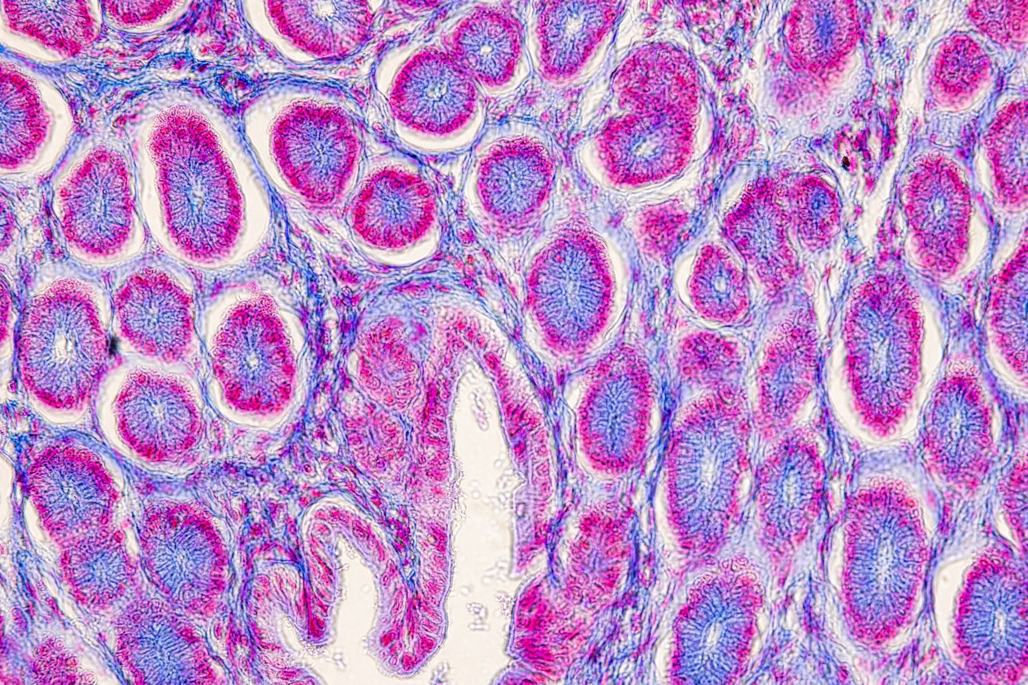
Image Credit: Sinhyu Photographer/Shutterstock.com
Protein leakage varies depending on several factors, including a protein’s subcellular location, physical properties, and interactions with other molecules. Because proteins are typically about ten times smaller than their corresponding messenger RNA (mRNA) templates, they’re more prone to leaking when cell membranes are compromised.
Recent advancements in single-cell proteomics using high-throughput mass spectrometry have made it possible to quantify proteins at cellular and even subcellular resolution. These measurements are crucial, as protein synthesis, degradation, abundance, and post-translational modifications all shape how individual cells function within tissues.
Minimizing changes to proteins during sample preparation, especially those related to tissue dissociation and storage, is key to preserving the integrity of the data. However, freezing samples can introduce cell damage, leading to biased results.
Similar issues have been observed in single-cell RNA sequencing (scRNA-seq), where transcripts may leak from permeable cells depending on their subcellular origin. In scRNA-seq workflows, permeabilized cells are often excluded using computational cutoffs tailored to each cell type. Until now, a comparable approach for proteomics has been lacking.
About the Study
To address this gap, the researchers designed a set of murine experiments to study protein leakage in mammalian cells.
They also developed a classifier that uses a specific protein leakage signature to identify permeabilized cells across different cell types and species. This classifier is integrated into a new tool called QuantQC, which enables researchers to detect and correct protein leakage artifacts, even without access to advanced sorting technologies.
For the experiments, the team used male C57BL/6 mice, euthanized via carbon dioxide followed by cervical dislocation, to isolate tracheal cells. A total of 2,784 cells were analyzed, either fresh (n=928) or after cryopreservation at -80 °C using a solution of 10 % dimethyl sulfoxide (DMSO) and 90 % fetal bovine serum (FBS) (n=1,856). To identify cells with compromised membranes, they stained the samples with Sytox Green, a membrane-impermeable dye, and defined the corresponding protein leakage signature.
The researchers trained an XGBoost classifier to distinguish permeabilized cells based on the abundance of the 75 most leakage-prone proteins. The model was validated using previously published human cell line data. It was trained on basal cells, immune cells, and fibroblasts, and tested on basal cells.
Using the prioritized Single-Cell Proteomics by Mass Spectrometry (pSCoPE) method, the team analyzed 1,018 cells per day, quantifying 712 proteins per cell via liquid chromatography-mass spectrometry (LC-MS). They calculated fold changes in protein abundance between permeable and intact cells, assigning cell types using the LIGER algorithm and reference single-cell mRNA datasets.
Key Findings
Sytox Green staining revealed that while 96 % of fresh cells remained intact, only 72 % of frozen cells did. The LIGER-based clustering showed that frozen samples had a higher presence of permeabilized cells, which grouped together and showed reduced alignment with expected transcriptomic profiles.
Most proteins were depleted in permeabilized cells, confirming significant leakage. This effect was especially pronounced for nuclear and cytosolic proteins, compared to mitochondrial or membrane-associated proteins. Among the most affected were cytosolic enzymes, including peroxidases and glycolytic enzymes like glyceraldehyde-3-phosphate dehydrogenase (GAPDH), which showed roughly two-fold decreases in abundance.
The XGBoost classifier performed well, achieving an area under the curve (AUC) of 0.92 when tested on the same cell type, and 0.86 when tested across different types. Notably, it showed strong consistency in leakage signatures between mouse and human cells, supporting its broad applicability.
Download your PDF copy now!
Conclusions
This study underscores the importance of accounting for protein leakage in single-cell proteomic analyses. By identifying and removing compromised cells—either through membrane staining or computational tools like QuantQC—researchers can improve the fidelity of their datasets and draw more accurate conclusions.
Future research could explore ways to mitigate protein leakage, such as using cross-linking reagents during sample preparation. A deeper understanding of how and why proteins leak could also offer new insights into cellular protein dynamics and stability.
Journal Reference
Leduc, A., et al. Limiting the impact of protein leakage in single-cell proteomics. Nat Commun 16, 4169 (2025). DOI: https://doi.org/10.1038/s41467-025-56736-7, https://www.nature.com/articles/s41467-025-56736-7