Trace Evidence and Microscopy
Trace evidence is an integral part of every forensic investigation, and valuable information can be extracted when they are analyzed correctly. Microscopy is a nano-scale technology capable of generating forensically relevant information and is widely used for almost any investigation within forensic science. There are many different types of microscopes, including light, electron, and probe microscopes. Their potential forensic applications include age determination of bloodstains, fingermarks, and gunshot residue analysis, among others.
A few commonly used light microscopes in forensics are the compound microscope, the polarizing light microscope, and the stereomicroscope. Electron microscopes, including Scanning Electron Microscopy (SEM) and Transmission Electron Microscopy, as well as probe microscopy such as Atomic Force Microscopy (AFM), are commonly used for forensic investigations.
Light microscopes utilize photons for image development, while electron microscopes take advantage of electrons for the same purpose. SEM and TEM, used in tandem with other analytical instruments (such as infrared spectroscopy), are currently the most commonly used tools for forensic analysis.
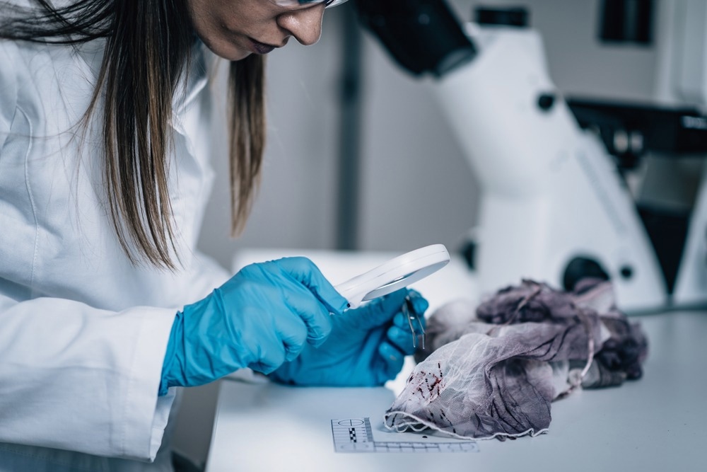
Image Credit: Microgen/Shutterstock.com
Examination of Soil Samples
Soil is commonly analyzed to provide evidence because soil particles may adhere to almost every item of forensic interest. Trace evidence/ particles found in soil can be quantitatively analyzed by SEM coupled with energy-dispersive X-ray spectrometry (SEM-EDS). A study by Kikkawaa et al. targeted the light minerals orthoclase albite, anorthite, and quartz to determine whether a statistically robust analytical method could discriminate between soil samples.
Minerals with specific gravities <2.9 are considered light minerals. Quantitative analysis by SEM-EDS is particularly suitable for light minerals because a sample will often contain far more light mineral particles than heavy mineral particles, meaning a statistically robust analysis can more easily be performed by utilizing light minerals instead of heavy ones.
Chi-squared tests were used for at least 100 particles per soil sample for the statistical analysis. Favorable outcomes were obtained using this methodology both on simulated and real soil samples across Japan.
Can Feathers Provide Forensically Relevant Information?
Feather evidence can sometimes be helpful to police casework in proving physical contact between clothing made by down feathers, or it may provide specific leads to the crime scene by identifying the species or sub-group of birds from which the feather evidence came from.
In their 2011 study, Dove and Koch created a guide that describes the importance of feather evidence to forensic investigations and introduces the basic techniques of approaching the identification of birds from feathers. Photomicrographs and descriptions are provided for multiple orders of birds commonly found in criminal cases with emphasis on the diagnostic microscopic features for each one.
Characteristics of feather barbs, barbules, nodes, and pigmentation patterns are described extensively with warning text for similar species in each group. Also discussed are details of feather topography, microslide preparation for downy feather barbs, information on report writing and testimony, and the general significance of feathers in forensic cases. Detailed instructions on how to study the feathers can also be found in the guide.
The authors suggest using a standard compound light microscope or comparison microscope with a magnification range of 40X400X and placing the cover slip over the feather barbs. The microslide should be allowed to dry before viewing. Viewing should be first performed at low power to get an overview of barb, barbule length, and pigment patterns. Viewing at high power is ideal for identifying node morphology details and diagnostic characteristics.
After following the protocol that the guide suggests, three conclusions can be reached from a microscopic examination of feathers: 1) Confirmation that feathers or feather portions are present in the submitted evidence. However, no diagnostic features present could lead to specific identification. 2) Identification of the order, family, or species of bird. 3) Identification of the order, family, or species of bird and comparison with a known source of the feathers. This can result in an association or exclusion of an item or bird.
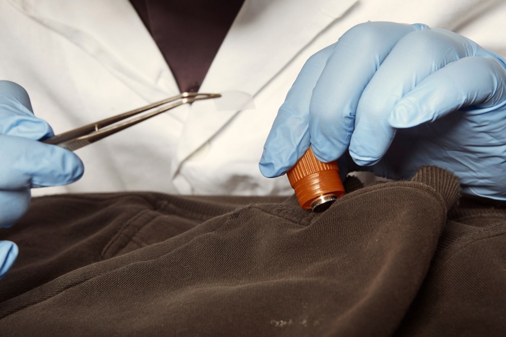
Image Credit: Couperfield/Shutterstock.com
AFM and its Multiple Uses in Forensics
Atomic force microscopy facilitates visualization and physicochemical surface characterization of materials at the nano-scale and sometimes at a molecular level. Multiple publications regarding bloodstains, fingermarks, textile fibers, document forgery, and gunshot residue have proved the usefulness of this nanotechnology in forensic investigations.
The greatest asset of minimally invasive AFM is its ability to measure material characteristics such as topography, morphology, elastic modulus, adhesion forces, energy dissipation, and dielectric properties. For forensic applications, investigation at this nanolevel does not necessarily benefit the overall aim of a forensic examination. When it comes to fingermark investigations, the roughness of the substrate may render exploration of the ridges) of the marks impossible. Surface roughness issues can be mitigated by allowing larger scan areas and adding extra phase imaging.
AFMs can image uneven surfaces as long as the roughness does not exceed the scanner's capacity in the vertical Z-direction, which usually ranges from 5 to 50 lm. If surface roughness exceeds this threshold, the scanning system will most likely crash.
Surface roughness is also responsible for unreliable, irreproducible force-distance curves when the probe is very sharp compared to the usual width. In this case, van der Waals and capillary forces from the sides of the AFM will also contribute to this effect. Tip-less cantilevers or spherical-shaped tips can rectify this problem, but it will be at the expense of resolution.
Choosing the Right Microscopic Technique
In actual police casework, microscopic techniques such as light microscopes and compound ones are ideal for hair analysis and identification due to their simplicity of use and low cost compared to other microscopy techniques such as SEM or TEM.
Transmission instruments (i.e., TEM) are generally more difficult to use and require more painstaking sample preparation than other microscopes, and they usually require the sample to be quite thin to facilitate the penetration of the electron beam. Scanning techniques (i.e., SEM) are a bit easier to use than transmission instruments, which is why it's generally preferred for the analysis of gunshot residue.
Continue reading about the various uses of microscopy in forensics here!
Further Reading
Last Updated: Nov 21, 2022