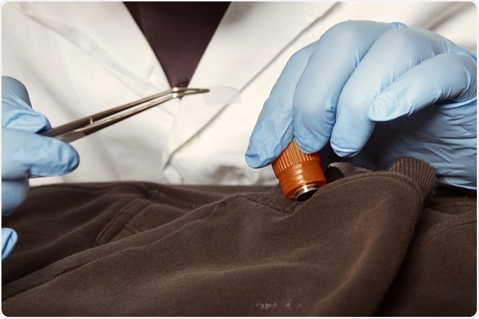
Forensic science has many applications in a variety of fields, forensic science can be used to investigate ancient civilizations and its primary use is within crime scene investigation in identifying evidence. Microscopy is vital to multiple areas within forensic science and there is a variety of techniques utilized.

Image Credit: Couperfield/Shutterstock.com
Microscopy in criminal science
Microscopy can be applied in the identification of trace evidence such as fragments, fibers, hairs, fingerprints which are left the crime scene, on a victim or suspect.
When a gun is fired, it leaves behind a chemical residue, known as gunshot residue (GSR). This residue can help identify a weapon used at a crime scene and link a suspect to a crime if the GSR is found to match that which was left at the scene of the crime.
The tiny particles that makeup GSR measure between a few hundred nanometers to a few microns.
Therefore, scanning electron microscopy and energy-dispersive X-ray spectroscopy (SEM/EDX) analysis may be used to investigate gunshot residue, distinguishing the shape of the particles and their chemical composition revealing their distinct chemical composition, allowing for the identification of a potential weapon.
Fragments of glass may be found in incidents such as house break-ins and road traffic accidents. Microscopes can be used to compare shattered glass left at the scene to that found on a suspect. Different types of glass have varying compositions, allowing scientists to analyze its origin by looking closely at its structure.
Fingerprints are incredibly useful for identifying perpetrators in a crime scene. Scanning electron microscopy and energy-dispersive X-ray spectroscopy can be used to magnify the prints and match them to a potential suspect or victim.
Microscopy in forensic epidemiology
Forensic epidemiologists investigate how disease spreads. In epidemiology, microscopes can be used to investigate the root of an outbreak by tracing the pathogen involved to its source. This information can be essential in preventing further infection, reducing the risk of an epidemic.
Microscopy in forensic anthropology
Microscopes have many uses in the field of forensic anthropology, the field of identifying the factors of death. For example, bone and other body tissues can be investigated using a microscope to gain clues about the cause of death.
Scanning electron microscopes are often used to study soil samples taken from the location of a body to determine the length of time the body had been there, the bones can be investigated for sharp force trauma and the coating left on the teeth may be examined, which can also be indicative of a cause of death.
Microscopy in forensic pathology
Similar to forensic anthropology, forensic pathology investigates and determines the cause of death. The difference between forensic anthropology and forensic pathology is that forensic pathologist aims to establish the exact cause of death, rather than indicators of potential events leading to death.
Microscopy can be used to identify the bacteria or virus which caused death, or to examine body tissue for wounds, determining cause and investigating the potential for the would be fatal.
Overall, there are a number of uses of microscopy in forensics. As technology continues to advance, it is expected that there will be further applications of microscopes in forensics.
Conclusion
Microscopy plays a vital role across multiple fields of forensic science, from crime scene investigation and forensic anthropology to pathology and epidemiology. By enabling the detailed examination of trace evidence, microscopic techniques help forensic scientists identify suspects, uncover the cause of death, and trace the origins of disease outbreaks. As microscopy technology advances, its applications in forensic science are expected to expand, further enhancing the accuracy and effectiveness of forensic investigations.
References:
- Bartelink, E. J., Wiersema, J. M., & Demaree, R. S. (2001). Quantitative analysis of sharp-force trauma: an application of scanning electron microscopy in forensic anthropology. Journal of Forensic Science, 46(6), 1288-1293.
- Jones, B. J., Downham, R., & Sears, V. G. (2010). Effect of substrate surface topography on forensic development of latent fingerprints with iron oxide powder suspension. Surface and Interface Analysis: An International Journal devoted to the development and application of techniques for the analysis of surfaces, interfaces, and thin films, 42(5), 438-442.
- Jones, B. (2019). Microscopy in Forensic Sciences. Springer Handbook of Microscopy, pp.2-2.
- Korda, E. J., MacDonell, H. L., & Williams, J. P. (1970). Forensic applications of the scanning electron microscope. J. Crim. L. Criminology & Police Sci., 61, 453.
- Meng, H. H., & Caddy, B. (1997). Gunshot residue analysis—a review. Journal of Forensic Science, 42(4), 553-570.
Further Reading
Last Updated: Nov 4, 2024