What is microscopy, and what are the different types used in healthcare?
The development has changed the visualization of cells and organisms in microscopy. Since their invention in 1600, different microscopic techniques, such as light and electron microscopy, have been used to study cells. These studies have evolved greatly from observing fixed cells to observing and generating quantitative data from live cells. The basic mechanism of a microscope involves an objective lens that allows the image to be magnified and the eyepiece. Different microscopes, such as optical microscopes, electron microscopes, fluorescence microscopes, digital microscopes, and others, can be used. This article will use examples to discuss how these techniques build the intersection between microscopy and healthcare.
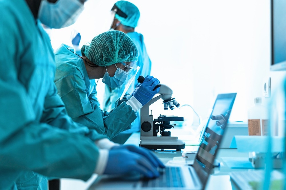
Image Credit: AlessandroBiascioli/Shutterstock.com
Use of microscopy in infectious diseases
To be able to diagnose, treat and understand different diseases, it is crucial to be able to visualize the pathogens/microorganisms at a molecular level. This is only possible due to the developments in microscopy. Light microscopy is one of the most common ways to visualize biological samples. However, conventional light microscopy, which is based on focusing a light beam on the object through a convex lens to enlarge the object, comes with the disadvantage that it cannot form a separate image of 2 objects closer together than 200nm. They form one image instead of 2 separate images, so more advanced imaging techniques have been developed, such as super-resolution imaging which can visualize images below this limit through fluorescence emission of only a specific set of molecules.
The development of this technology has created breakthroughs in healthcare by allowing the observation of interactions between hosts and pathogens, such as HIV, in detail at a nanometer scale. It is also possible to view the pathogens at a molecular level, such as the HIV particle budding from cells.
Although the resolution of the images is still not at the level of electron microscopy (EM), it allows researchers to investigate questions previously reserved only for EM. However, the development of EM is still the main way to detect pathogenic organisms such as viruses and proves to be a powerful diagnostic tool in the healthcare field, even leading to the discovery of new viruses. EM is advantageous in diagnostics as it doesn't depend on prior knowledge about the virus, unlike serological methods, which need a specific probe to identify the virus.
Recent advances that build the intersection between microscopy and healthcare
As mentioned, microscopy has been fundamental to diagnosing diseases and is even more important during major healthcare crises such as the SARS-CoV-2 pandemic. EM was used to isolate and identify the structural characteristics of SARS-CoV- 2, which allowed healthcare professions to be better informed about the virus and carry out further research.
However, advanced technologies such as high-throughput microscopy were also used during the pandemic. The advantage of this technique is that it uses biochemical assays, which allow in-vivo models to be developed, which show, more accurately, how drugs behave in terms of metabolism and toxicity.
During the SARS-CoV-2 pandemic, fluorescent proteins were inserted into the genome to screen for anti-viral drug compounds and other research. A successful example of this was a research study in which a green fluorescent protein was inserted into the SARS-CoV-2 genome, causing a partial deletion in the ORF7a protein, which reduced SARS-CoV-2 replication, and another study observed the opposite effect where insertion of the same protein cause increased replication.
Another breakthrough after EM in the field of microscopy is cryo-electron microscopy (cryo-EM). Cryo-EM can produce the highest quality images and has also been able to identify individual atoms. This has allowed researchers to understand in detail how proteins work. This would allow drug companies to visualize protein structures to develop drug interaction models, allowing for more accurate drug development.

Image Credit: cono0430/Shutterstock.com
How can deep learning (DL) be used in microscopy to aid healthcare research?
Although these advancements have revolutionized the intersection between microscopy and health care, limitations exist when obtaining clear images of biological specimens. For example, it can be difficult to ascertain a balance between the toxicity of fluorescent labels and getting a good image resolution. Additionally, images usually come with large datasets that need to be analyzed to identify useful information, which can require long hours of manual extraction and analysis. This may cause issues such as human error, which impacts the information's reliability and affects diagnostics and research.
A new field of research known as Deep Learning (DL) provides new avenues in healthcare and microscopy that tackle these limitations. DL is a section of artificial intelligence (AI) based on recognizing patterns to predict outcomes in unfamiliar data, and DL relies on neural networks (NN), which can be trained in a process known as inference. Inference uses a 'training dataset', i.e., a known input of data that results in the output. Based on this training dataset, the NN can learn to analyze completely unknown data sets to provide an output.
NN can be used in microscopy to identify and classify images that aid diagnosis. Research has shown that NNs can detect cancer without the manual labor of differentiating between relevant image features.
An example of this was seen in a study that used NNs to detect mitosis in images taken of breast cancer tissue samples, which is important for screening and assessing the progress of cancer.
The field of microscopy is evolving, and the intersection between this ground-breaking technology and healthcare is always being improved and investigated. With the advent of technologies such as DL and NN, healthcare interventions to diagnose and treat diseases are improving and becoming more reliable and accurate and will continue to do so.
Sources:
- Callaway, E. (2020). ‘'It opens up a whole new universe': Revolutionary microscopy technique sees individual atoms for first time' Nature, 582 (7811), pp. 156-157. DOI: 10.1038/d41586-020-01658-1.
- Chen, X., Zheng, B. & Liu, H. (2011). 'Optical and digital microscopic imaging techniques and applications in pathology' Anal Cell Pathol (Amst), 34 (1-2), pp. 5-18. DOI: 10.3233/acp-2011-0006.
- Cireşan, D. C., Giusti, A., Gambardella, L. M. & Schmidhuber, J. (2013). 'Mitosis detection in breast cancer histology images with deep neural networks' Med Image Comput Comput Assist Interv, 16 (Pt 2), pp. 411-8. DOI: 10.1007/978-3-642-40763-5_51.
- Laketa, V. (2018). 'Microscopy in infectious disease research-imaging across scales' J Mol Biol, 430 (17), pp. 2612-2625. DOI: 10.1016/j.jmb.2018.06.018.
- Reigoto, A. M., et al. (2021). 'A comparative study on the use of microscopy in pharmacology and cell biology research' PLoS One, 16 (1), p. e0245795. DOI: 10.1371/journal.pone.0245795.
- von Chamier, L., Laine, R. F. & Henriques, R. (2019). 'Artificial intelligence for microscopy: What you should know' Biochem Soc Trans, 47 (4), pp. 1029-1040. DOI: 10.1042/bst20180391.
Further Reading
Last Updated: Jan 17, 2023
 Supramolecular Shampoo Uses Self-Assembly to Replace Polymer Thickeners