This loss of tension may serve as a mechanical biomarker linked to tumor grade. Tumor grading is essential for diagnosis and treatment, as it helps predict aggressiveness, treatment response, and recurrence risk.
The results suggest that targeting physical changes in extracellular matrix (ECM) fibers could offer new therapeutic options for breast cancer.
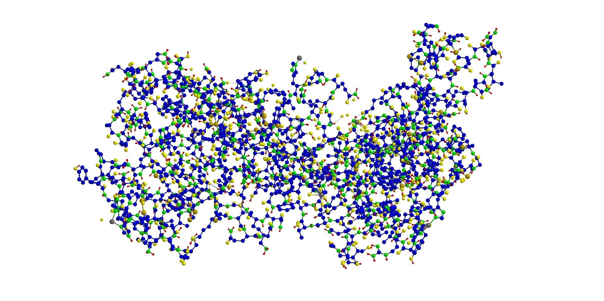 Molecular structure of Fibronectin. Image Credit: Raimundo79/Shutterstock.com
Background
Breast carcinomas are among the most common cancers globally. ECM remodeling, involving structural and compositional changes, plays a major role in cancer progression. This remodeling is driven by interactions between the ECM and contractile cancer-associated fibroblasts (CAFs).
Cells apply mechanical forces to ECM fibers, altering their tension. These changes can affect the availability of growth factors and cytokines bound to the ECM, which are important for cell survival. Fibronectin is a key ECM protein involved in cell-ECM interactions that influence tumor behavior. However, how fibronectin tension changes during cancer development is not fully understood.
About the Study
Researchers used Cy5-FnBPA5 peptide probes to study fibronectin fiber tension in cancer. These probes bind to relaxed (non-tensed) fibronectin fibers with high affinity. As fiber tension increases, the probe’s binding affinity decreases.
In a mouse tumor model, they observed relaxed fibronectin fibers in tumor tissue. In contrast, fibronectin fibers in healthy organs were under high tension. In tumors, fibronectin and collagen fibers were located near CAFs marked by α-smooth muscle actin (α-SMA). Regions with relaxed fibronectin also showed the presence of tenascin C (TNC) and immune cells with immunosuppressive properties.
Based on these mouse findings, the team investigated whether human breast cancer tissues also show relaxed fibronectin fibers and how this affects the tumor microenvironment.
Human Tissue Analysis
Tissue samples were collected from healthy breast tissue and breast cancer cases through the Kantonsspital Baden (KSB) and the Tissue Bank Bern (TBB) in Switzerland.
Healthy samples came from breast reduction surgeries or from non-cancerous areas near tumors. Tumor samples included ductal carcinoma in situ (DCIS) and invasive breast cancer. None of the patients had received chemotherapy or radiation before surgery.
Tissues were analyzed using immunofluorescence, confocal microscopy, and multi-photon imaging. The team examined markers such as GATA3, pan-cytokeratin (panCK), Ki67, CD34, DAPI, and TNC.
Collagen fiber arrangement was assessed through second harmonic generation (SHG) imaging and spatial analysis. Proximity analysis measured the distance between fibronectin fibers and cancer or immune cells. A ratiometric approach compared Cy5-FnBPA5-positive signals to total fibronectin to distinguish relaxed from tensed fibers.
Results
Fibronectin fibers remained tensed in healthy breast tissues and DCIS but appeared relaxed in invasive tumors, mirroring results from the mouse models. Confocal microscopy showed a fibrotic ECM in invasive cancers, with strong TNC and SHG signals, and increased FnBPA5 binding.
FnBPA5 signal intensity rose by 200 % in high-grade tumors compared to intermediate ones. In contrast, DCIS tissues showed weaker fibrotic signals and limited FnBPA5 binding, mostly around glands and ducts. Healthy tissues lacked SHG, TNC, and FnBPA5 signals.
Healthy tissue had wavy, loosely organized collagen fibers forming a structural network around ducts and glands. Invasive tumors showed collagen fibers arranged linearly and perpendicular to ducts, pulled by CAFs invading the surrounding stroma. DCIS retained collagen and fibronectin organization similar to that of healthy tissue.
Quantitative image analysis confirmed more relaxed fibronectin fibers (FnBPA5+ pixels) in invasive tumors than in DCIS or healthy tissue. Collagen structure also varied significantly across the three tissue types.
Healthy tissues featured GATA3+ and panCK+ cells arranged in acinar structures, with few Ki67+ proliferative cells located away from relaxed fibronectin. α-SMA+ cells were found near CD34+ vessels, indicating smooth muscle cell origin.
In contrast, invasive cancer tissues had disorganized GATA3+ and panCK+ cells. Many Ki67+ cells with dense nuclei were located near relaxed fibronectin fibers, within 11 μm of FnBPA5+ signals. These tissues also had fewer α-SMA+ cells, located near relaxed fibronectin but farther from CD34+ vessels, suggesting a CAF origin. Over 60 % of T cells in the tumor stroma were located near relaxed fibronectin.
DCIS samples showed features between those of healthy and invasive tissues.
Download your PDF copy now!
Conclusion
The study suggests that fibronectin fiber relaxation is associated with breast cancer progression. Relaxed fibronectin fibers may represent a mechanical and chemical signature of invasive tumors and could help differentiate tumor grades or guide new therapies.
Journal Reference
Miéville, A, et al. (2025). Fibronectin Fibers Progressively Lose Their Tension in Invasive Human Breast Carcinoma while Being Tensed in DCIS and Healthy Breast Tissue. Adv. Sci. DOI: 10.1002/advs.202404351, https://advanced.onlinelibrary.wiley.com/doi/10.1002/advs.202404351
 New Protein Storage Condensates May Hold Clues for Better Anticancer Strategies