Our exposure to any type of radiation, whether that be natural radiation present within the earth or radioactive particles that have been deposited after a nuclear accident, can lead to long-term health effects like cancer and cardiovascular disease. The identification of these radioactive particles present within our environment is therefore critical and can be achieved by several different analytical techniques.

Image Credit: fewerton/Shutterstock.com
Source of radioactive particles
Within the environment, radioactive particles often originate from background radiation, which is present on Earth at all times. One type of background radiation is cosmic radiation, which is emitted from high energy particles, like the sun and the stars, and released into the Earth’s atmosphere. Whereas some cosmic radioactive particles can reach the ground, others will interact with the atmosphere to create different types of radiation.
Another type of background radiation can originate from naturally occurring minerals in the soil, ground, and water. Terrestrial radiation, for example, is derived from primordial radionucleotides like uranium and thorium and can be found in varying concentrations throughout the earth. Notably, surface soils often have the highest concentrations of both uranium and thorium.
The final type of background radiation is that which is emitted from human activities. For example, nuclear weapon tests and accidents, such as the Chernobyl disaster of 1986 that occurred in Ukraine, as well as nuclear reactors and industrial radioactive materials can all contribute to a small fraction of background radiation.
The behavior of particles in the environment
Regardless of their source, radioactive particles can be deposited into different types of ecosystems in several different forms including low molecular mass (LMM) species, colloids, nanoparticles, pseudocolloids, or fragments, to name a few. In addition to their form, other particle characteristics that can influence their behavior within the environment include particle size, crystalline structure, and oxidation states.
Once deposited into a given environment, several different interactions and transformation processes can cause the dispersion and growth of the particles to change. These processes can range from weathering rates and mobility to the biological uptake of these particles into the environment. As the inherent properties of the radionuclides are altered, their original distribution within a given environment subsequently changes as well.
Contaminated soils and sediments, for example, can act as transient sinks for radionuclides by allowing these particles to collect here until they are eventually transferred to the water phase through complexation with organic compounds or reduction and oxidation processes.
Other factors that influence the environmental behavior and biological effects of radioactive particles include how they were released into the environment and the inherent conditions of the ecosystem in which they have been deposited.
Radioactive particle characterization techniques
The most common types of analytical techniques that have been applied to the characterization of radioactive particles include in situ fractionation, solid-state non-destructive techniques, semi-destructive speciation techniques as well as several different methods involving a full decomposition of particles.
In situ fractionation techniques
Traditional approaches to collect radioactive particles that are transported by air have involved the use of air filter devices; however, cascade impactors have become increasingly used for this purpose over the past several years.
The advantage of using a cascade impactor is that particles of different aerodynamic diameters can be collected at the same time and placed on different impactor stages that represent a given size class. Samples can then be taken from a given impactor stage and analyzed by several different techniques such as radioanalysis and/or mass spectrometry (MS).
Comparatively, the measurement of radioactive particles within water sources requires the incorporation of both size and charge fractionation techniques. Some of the more frequently applied in situ techniques for this type of sample medium include filtration, which includes membrane sizes of 0.45, 0.2, and 0.1 micrometers (µm), and ultrafiltration, which has membrane sizes within the range of nanometers (nm) to µm.
When these size fractionation techniques are combined with charge fractionation methods, such as cation chromatography, radionuclides can be separated by both charged or neutral species, as well as colloids or particulate materials. This combination of size and charge fractionation techniques is often followed by radioanalytical and/or MS techniques.
Several different analytical techniques can measure the extent of particle heterogeneity present within a sample. Certain radiation-based imaging techniques like autoradiography, beta-camera, alpha track analysis, and fission track analysis can not only measure heterogeneities but can also provide quantitative information on the distribution and migration of radioactive particles.
Repeated sample splitting combined with an imaging technique like light microscopy, g-spectrometry or autoradiography can also be used to measure heterogeneities.
Solid-state non-destructive techniques
Once a sample has been obtained, both hot spot identification, which is achieved by phosphorus imaging, as well as sample splitting is conducted before particle identification. Micromanipulators can then be used under a light microscope to extract specific amounts of the sample that can even be in the form of single-particle extractions.
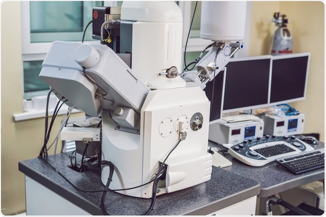
Particle characterization can then be conducted in various ways, depending upon what information must be obtained. Scanning electron microscopy (SEM) combined with energy dispersive X-ray analysis (EDX), for example, can provide information on particle size distribution, surface morphology, and the composition and distribution of elements present on the particle surface.
The combination of either SEM or ESEM with EDX is often used as a preparative method to indicate whether more advanced nano- or microanalytical techniques, such as laboratory-based, or synchrotron radiation-based X-ray techniques are needed.

Image Credit: Elizaveta Galitckaia/Shutterstock.com
Advances in Sccaning Electron Microscopy
References and Further Reading
- “Radiation Sources and Doses” – United States Environmental Protection Agency
- Salbu, B., & Lind, O. C. (2020). Analytical techniques for characterizing radioactive particles deposited in the environment. Journal of Environmental Radioactivity 211. doi:10.1016/j.jenvrad.2019.106078.
- Oki, Y., Tanaka, T., Takamiya, K., Osada, N., et al. (2016). Size Measurement of Radioactive Aerosol Particles in Intense Radiation Fields Using Wire Screens and Imaging Plates. Journal of Radiation Protection and Research 41(3); 216-221.
Further Reading
Last Updated: Oct 1, 2020