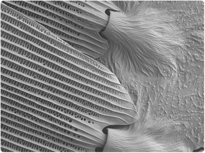
What is SEM?
Scanning electron microscopes, known as SEMs, have been used for close to a century. In 1937, Manfred Von Ardenne developed the scanning transmission electron microscope. He developed an advancement of the classic microscope that enabled scientists to view specimens in much greater detail.

Image Credit: CaludiaSEM/Shutterstock.com
In SEM, an electron beam replaces the light beam used in conventional microscopes to reveal high-resolution, three-dimensional images that unveil information on the sample’s topography, morphology, and composition.
The technology relies on electromagnetic converging lenses that control the path of the electron beam and the condenser that controls the resolution of the image produced by determining the size of the beam.
Upon interaction with the electron beam, the sample emits different types of electrons. The Backscattered Electron (BSE) detector can pick up these backscattered electrons.
The images produced by the SEM demonstrate areas of different chemical compositions through the differing contrast generated.
Elements with a higher atomic number are shown as brighter, and those with lower numbers are darker.
More detailed information is obtained by the SEM’s Secondary Electron (SE) detector that is positioned adjacent to the electron chamber, increasing the efficiency of detecting secondary electrons.
Applications of SEM
Many industries have adopted SEM into their procedures. Commercial, scientific, medical, industrial, and research sectors all heavily rely on the SEM, which helped them to develop their methodologies and equipment. Below is an overview of how different sectors are currently using SEM.
Materials science is using SEM for research, quality control, and failure analysis. Research into nanotechnology is also relying on this technology to help explore how nanowire can improve sensor technology.
It is also being used to enhance the performance of newly developed semiconductors, testing their efficacy and visualizing their topographical structure.
The electronics sector has greatly benefitted from the use of SEMs which have been used to deepen our knowledge of the best practices for producing microchips.
With the Internet of Things (IoT) rapidly infiltrating every corner of our daily lives, the use of SEMs will become increasingly important in this field, as the need grows to gain insight into how to produce cheaper, lower-power electrical components.
Several applications of SEMs have arisen in the field of forensic science. SEMs are currently relied upon for the tasks of handwriting and print analysis, gunshot residue analysis, bullet marking comparison, jewelry examination, fraudulent document analysis, paint particle, fiber analysis, filament bulb analysis in traffic incidents, and more.
Perhaps the areas that have benefited the most from the use of SEMs are biological science and medical science.
Here, SEMs are used in both research and clinical applications to identify new bacteria and strains of virus, monitor the impact of climate change, test new vaccines, uncover new species, assist in genetic testing, identify new diseases, test new medicines, compare tissue samples. SEMs also help gain insight into the nature of various diseases, how they establish, progress and respond to treatment.
Recent advances
This rise in the use of SEM in the life sciences has been attributed as the main driving force for the advancement in SEM technology.
While SEM has not drastically changed in terms of design and concept since it was first introduced in the 1930s, its resolution and power have been improved over the years.
However, due to its use in the biological sciences, SEMs have gone through significant advances in recent years to support its role in this field. Since the early 2000s, the use of SEM has rapidly increased, with the number of publications referring to the method growing year on year.
One area in particular that has pushed advancement in this technology is neuroscience. The discipline has begun developing SEM to help solve previously unaddressable problems. Scientists have applied SEM to help uncover the physiological changes occurring in the central nervous system, leading to recent advances in the technology and function of SEMs, such as the establishment of serial block-face SEM (SBEM) which is used to image blocks of ultra-thin tissue removed using an ultramicrotome that is then incorporated into the electron microscope.
This technology produces three-dimensional images of neural tissue, allowing scientists to explore neurons at an unprecedented level of detail.
The development of FIB-SEM (Focused Ion Beam Scanning Electron Microscopes) represents another major advancement in SEM technology that has been born out of its use in life sciences.
In FIB-SEM, a focused beam of ions replaces the use of a beam of electrons.
In this method, the ion beam is used to remove ultrathin layers of tissue, allowing the reconstruction of z-stacks.
The major benefit of the FIB-SEM method is that it can produce resolutions of up to a few nanometers, enabling the visualization of the intracellular environment in high resolution.
Finally, there have also been numerous general advances made to the hardware used in SEM over recent years. For example, the lenses and detectors have been improved to allow the resolution power of the SEM to increase.
Specifically, scanning columns for electron spectrometers have advanced so that they can now be placed inside a cylindrical mirror energy analyzer; testers and lithographs for semiconductor technology; and miniaturized versions.
Simultaneous to these advancements in SEM has been the developments seen in protocols for scientific procedures, such as those in immunolabeling, preservation of the ultrastructure, imaging of the cytoskeleton and the inner structures of host cells, and more.
This demonstrates the importance of SEM technology in the biological sciences, and how advances in this technology coincide with advances in its applications.
Sources:
- de Souza, W. and Attias, M. (2018). New advances in scanning microscopy and its application to study parasitic protozoa. Experimental Parasitology, 190, pp.10-33. www.sciencedirect.com/.../S0014489417305829?via%3Dihub
- Frank, L. (2002). Advances in scanning electron microscopy. Advances in Imaging and Electron Physics, pp.327-373. https://www.sciencedirect.com/science/article/pii/S1076567002800696
- Shi, C., Luu, D., Yang, Q., Liu, J., Chen, J., Ru, C., Xie, S., Luo, J., Ge, J. and Sun, Y. (2016). Recent advances in nanorobotic manipulation inside scanning electron microscopes. Microsystems & Nanoengineering, 2(1). https://www.nature.com/articles/micronano201624
Further Reading
Last Updated: Mar 20, 2020