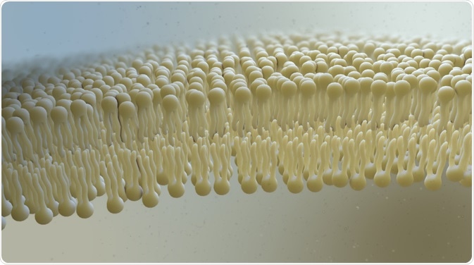
Lipids are molecules that have a strong propensity to associate with one another through non-covalent forces and are characterized by having a hydrophilic head group and a hydrophobic tail group. Biological lipids are insoluble in water and tend to aggregate due to non-covalent bonding.
The major types of lipids are simple fatty acids, triglycerides, phospholipids, sphingolipids, sterols (such as cholesterol), carotenoids, and prostaglandins. Raman spectroscopy is a technique that detects vibrational motions in molecules and can be used when studying lipids. It is a powerful technique and often used when analyzing biological samples such as lipids and proteins because it’s non-invasive, highly accurate, and provides abundant chemical information.

Image Credit: Nathan Devery/Shutterstock.com
Lipids in the body
Fatty acids consist of a carboxylic acid head group with a long unbranched aliphatic tail. The long hydrocarbon tail is either saturated with hydrogens or unsaturated (containing double bonds). They are found in natural fats and oils and as a component of membrane phospholipids and glycolipids. Fatty acids are generated by the hydrolysis of the ester linkages in triglycerides liberating glycerol and fatty acid.
Triglycerides (also termed triacylglycerols (TAGs) or neutral fats) are composed of three fatty acids each in ester linkage with a single glycerol molecule. This results in the polar hydroxyls of glycerol and the polar carboxylates of the fatty acids being bound in the ester linkages, which causes TAGs to be nonpolar. TAGs have a significant advantage over polysaccharides (such as glycogen and starch) as a source of stored fuel. Stored TAGs are easily hydrolyzed by enzymes (lipases) found in lysosomes to give fatty acids and glycerol; the former is further broken down after they are carried to target tissues via the bloodstream.
Other common lipids in biological systems are sphingolipids, which are found most commonly as sphingomyelins (ceramide phosphocholines) in the nervous tissue and brain of humans and are important in the normal functioning of the nervous system. Sterols (such as cholesterol) are present in the cell membranes of most eukaryotic cells and play important roles in the synthesis of biological molecules, including bile acids, vitamin D, cortisol, and sex hormones such as estrogen and testosterone.
Carotenoids are lipid-soluble photosynthetic pigments made up of isoprene units and function to absorb photons (light energy) for use in photosynthesis while protecting chlorophyll from photo-degeneration. Prostaglandins are lipid-soluble, hormone-like regulatory molecules containing a five-carbon ring originating from the chain of arachidonic acid. They act as mediators and have complex effects on metabolism.
Phosphoglycerides are the major class of phospholipids and are the main component of biological membranes. Phospholipids are found in myelin sheaths surrounding axons and are found as granules in blood platelets (thrombocytes).
In biospectroscopy, phospholipids are most commonly studied in the form of phospholipid membrane bilayers. Phosphoglycerides are glycerol-based phospholipids and are derived from glycerol-3-phosphate. They are characterized by a phosphate head group attached in a glycerol unit to two acyl chains. In general, they contain a C16 or C18 saturated fatty acid at C-2 (2nd carbon position) and a C18 to C20 unsaturated fatty acid at C-3 (3rd carbon position). The cis stereochemistry of the alkene in the unsaturated fatty acid chain causes the chain to kink and when packed in a parallel fashion to form extended membrane sheets, the fluid mosaic property of the membrane is increased.
Phospholipids do not cross the membrane but rather move in lateral directions along the membrane while proteins never rotate in the membrane and remain in a fairly fixed position. There are several vibrational spectroscopy techniques used to analyze these biological lipids found in the body. However, Raman spectroscopy is the most non-invasive and can be used with aqueous samples.
Raman Spectroscopy of lipids
The industry standard for imaging biological material is using fluorescence spectroscopy, which is poor at imaging lipids due to the lack of fluorescence. Furthermore, mass spectroscopy can be used, however, due to the destructive nature of samples, it may struggle to speciate different forms of lipids. In contrast, Raman spectroscopy is non-destructive and is often used for understanding lipids in biological samples.
Raman spectroscopy is a spectroscopic technique used to study vibrational motions in molecules. It is a complementary technique to infrared spectroscopy. However, Raman spectroscopy is a scattering technique, while infrared spectroscopy is an absorption technique. The vibrational assignment of phospholipids and TAGs is based on several infrared studies that have investigated model lipids and biological membranes.
However, since Raman and infrared spectroscopies are complementary techniques, the band assignments of lipids can be equivalent. The non-polar nature of lipids gives it a very strong Raman cross-section due to the highly polarizable C-H and C-C bonds of the hydrocarbon chain.
It is also very common to use multivariate statistical techniques combined with Raman spectroscopy to detect biological samples such as lipids. These statistical techniques can help understand the physicochemical properties of lipids and help characterize them. In addition, predictive techniques such as partial least squares regression (PLSR) has been used to detect the concentration of lipids in cells and biological samples.
Sources:
- Matthews, C. K.; van Holde, K. J. Biochemistry; The Benjamin/Cummings Publishing Company: Redwood., USA, 1990.
- Lehninger, A.; Nelson, D. L.; Cox, M. M. Principles of Biochemistry, 3 ed.; Worth Publishers: New York, 2000.
- Astbury, W. T.; Dalgliesh, C. E.; Darmon, S. E.; Sutherland, G. B. B. M. Nature 1948, 162, 596-600.
- Mantsch, H. H.; McEthaney, R. N. Chem. Phys. Lipid 1991, 57, 213-226.
- Schultz C, Neef AB, Gadella TW, Goedhart J. 2010Imaging lipids in living cells. Cold Spring Harbor Protocol. 2010
- Passarelli MK, Winograd N. 2011Lipid imaging with time-of-flight secondary ion mass spectrometry (ToF-SIMS). Biochimica et Biophysica Acta (BBA) Mol. Cell Biol. Lipids 1811, 976-990
Further Reading
Last Updated: Aug 17, 2021