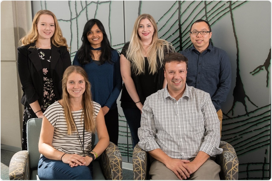
A team of researchers from IU School of Medicine is using human stem cells to study degeneration in glaucoma. Image Credit: Indiana University School of Medicine.
The scientists discovered that when differentiating pluripotent human stem cells into retinal ganglion cells, they were able to detect the traits related to neurodegeneration in glaucoma.
Once you’ve identified a target like this—what’s going wrong in the cells—this opens up a number of possibilities for the eventual development of therapeutic approaches, especially pharmacology approaches to slow down and reverse these degenerative phenotypes.”
Jason Meyer, PhD, Associate Professor, Department of Medical and Molecular Genetics, Indiana University School of Medicine
The scientists were headed by Meyer, together with the co-first authors of the publication, Kirstin VanderWall, and Kang-Chieh Huang, both graduate students from the School of Science at IUPUI in Meyer’s laboratory, which is situated within Stark Neurosciences Research Institute. Earlier, Meyer’s laboratory was situated within the School of Science.
When retinal ganglion cells degenerate as a result of glaucoma, it results in loss of vision and ultimate blindness. In this study, the scientists acquired pluripotent stem cells from a patient who had a genetic form of glaucoma, Meyer stated. Later, they differentiated the stem cells into retinal ganglion cells to look for neurodegeneration deficits.
One of the powerful things about (stem cell research) is when you get the cells from a patient that has a genetic basis for a disease; all of the blueprints are there in the cell’s DNA to develop features of the disease.”
Jason Meyer, PhD, Associate Professor, Department of Medical and Molecular Genetics, Indiana University School of Medicine
The team also employed gene-editing technology—called CRISPR-Cas9—to introduce a genetic mutation often related to glaucoma into the existing stem cell lines for disease modeling, and also to rectify the defective gene in patient-derived cells.
According to Huang, “CRISPR/Cas9 gene-editing approaches not only allowed us to study the disease but using this approach we were also able to show how correcting the gene mutation reversed the disease, demonstrating the potential for gene therapy approaches as well.”
Meyer added that the researchers found dysfunction in the autophagy process, the body’s way of eliminating damaged cells to regenerate healthy ones.
“We found that in the glaucoma patient cells, there are some deficits in this autophagy process, so you had too much cellular junk that was being built up,” Meyer stated, adding that those deficits corresponded with the degeneration of the cells, which would shrivel up and die ultimately.
With the help of a pharmaceutical compound known as rapamycin—recognized to enhance the autophagy process—the researchers found that several of the neurodegenerative traits earlier identified by them had slowed down and the cells appear to recover and seem more normal, added Meyer.
Meyer also stated that human stem cells are instrumental in analyzing human disease, particularly neurodegeneration. Previous studies on retinal ganglion cells and glaucoma as a degenerative disease using animal models imply variations in how cells react between species.
Since they are human cells, it gives somewhat of a more representative model for us to test pharmacological compounds, and it gives us a better idea of how it could potentially be toxic or nontoxic to human cells compared to testing compounds in animals.”
Kirstin VanderWall, Study Co-First Author, Indiana University School of Science, IUPUI
Having detected a target within the cells—the process of autophagy—the ongoing work of the laboratory will continue to analyze ways to apply various types of pharmaceutical compounds for glaucoma treatment, added Meyer. As is the case for several neurodegenerative diseases, like Parkinson’s and Alzheimer’s diseases, there are very few treatments, if any, and no remedy.
“There is a dire need to try and identify new approaches to treat these diseases,” Meyer concluded.
Source:
Journal reference:
VanderWall, K. B., et al. (2020) Retinal Ganglion Cells with a Glaucoma OPTN(E50K) Mutation Exhibit Neurodegenerative Phenotypes when Derived from Three-Dimensional Retinal Organoids. Stem Cell Reports. doi.org/10.1016/j.stemcr.2020.05.009.