In a recent study published in Advanced Functional Materials, researchers investigated tissue preference, or tropism, in the development of endometriosis. They identified ovarian-specific extracellular matrix (ECM) attachment motifs that increase endometriotic cell attachment. These motifs, particularly combinations containing collagen I, fibronectin, and laminin, promote stronger adhesion in endometriotic cells, especially on stiffer substrates that mimic fibrotic ovarian tissues.
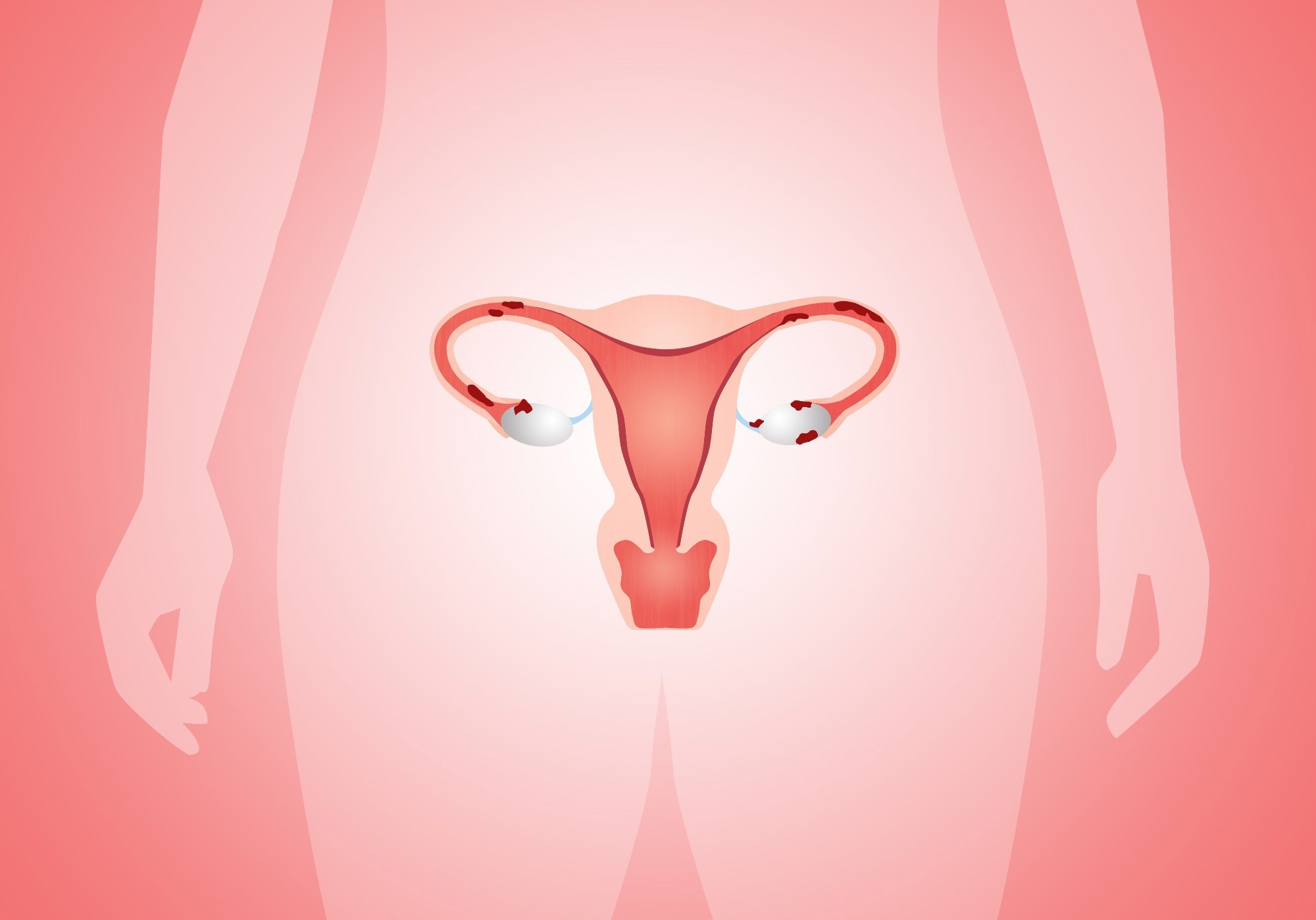 Image credit: Sogno Lucido/Shutterstock.com
Endometriosis is a chronic inflammatory condition affecting a significant number of menstruating females and those facing infertility. The disease involves the growth of tissues similar to the uterine lining in ectopic or non-native sites, often found on pelvic organs, particularly the ovaries.
Endometriotic lesions hamper assistive reproductive therapeutic efforts and can cause pelvic pain, dysmenorrhea, and menorrhagia. The initiation of endometriotic lesions remains unclear, as clinicians usually diagnose endometriosis at a late stage. Improving the understanding of mechanisms underlying the initiation of endometriosis can facilitate early diagnosis and timely medical attention, potentially improving fertility outcomes and quality of life.
Current theories, such as retrograde menstruation, cannot explain lesion formation, as it occurs in most menstruating individuals. Ablation procedures during diagnosis can also lead to fibrotic events, complicating the understanding of lesion initiation.
About the study
In the present study, researchers investigated the impact of ECM tropism on initiating endometriosis. They hypothesized that abnormal expression or function of cell-matrix (integrins) and intercellular (cadherins) adhesion molecules in the ovarian tissue microenvironment could contribute to the initiation and progression of endometriosis and ovarian cancer.
Polyacrylamide (PA) microarray systems with defined mechanical stiffness identified tissue-specific ECM and attachment motifs that affect endometriotic cells' size, morphology, and attachment. T-human endometrial stromal cell (ESC) and endometrial epithelial cell (EEC) cultures represented the native endometrium. The immortalized 12z human endometriotic epithelium-like and ectopic endometrial stromal cell (iEC-ESC) lines represented endometriotic lesions.
ECM and attachment factor biomolecules (250 μg/mL) on tissue culture plates enabled microprinting ECM combinations. Different stromal and epithelial cells seeded onto the microarrays created endometriotic and non-endometriotic cohorts for co-culture analysis.
The researchers determined the impact of cellular origin (endometriotic versus non-endometriotic), substrate stiffness (1.0 to 6.0 kPa), and multicellular (epithelial and stromal) cohorts to identify ovarian-specific adhesion motifs that increase endometriotic cell adhesion. Based on their actin protein distribution, they assessed eccentricities and distribution of endometriotic and non-endometriotic epithelial and stromal cells on adhesive islands 24 hours after culture. They investigated adhesion mechanisms of endometriotic epithelial (EndoE) and endometriotic stromal (EndoS) cells. The controls comprised liver tissues without endometriotic lesions. Statistical analysis using z-scores and linear regression models enabled quantitative comparisons across conditions.
Results
The team observed stronger endometriotic cellular adhesion on stiffer gels (6.0 kPa) than on softer gels (1.0 kPa) for all cells except non-endoE cells. The stiffer substrate resembles fibrotic or older ovaries. A stiff matrix generates positive feedback, potentiating fibrotic alterations within endometriotic cells through collagen I accumulation, increased proliferation, and cell differentiation. The findings suggest that therapies altering tissue stiffness or inhibiting fibrotic processes could improve disease prognosis.
The study demonstrated significantly elongated and wider EndoS cells than endoE and rounded non-endoE cells. Cell traction, mechanical forces that cells generate upon interaction with the ECM and surrounding cells, positively correlates with cell surface area, indicating increased adhesion in endoS cells, characteristic of myofibroblasts associated with endometriosis.
EndoE cells showed significantly increased attachment and eccentricity on the 6.0 kPa co-culture islands compared to non-endometriotic cells, aligning with the disease state, especially among cells undergoing epithelial-mesenchymal transition (EMT), associated with increased invasiveness. Changes in cell morphology (e.g., increased eccentricity and altered cell area) observed in endometriotic cells, particularly in co-cultures, may serve as prognostic indicators for disease severity or invasiveness. The findings suggest that monitoring the cellular characteristics could help predict disease progression or recurrence.
Collagen I and fibronectin were among the ECM components most significantly promoting endometriotic cell adhesion on stiffer substrates. Both proteins promoted the adhesion of endoS cells across various matrix combinations of laminin, a protein elevated in the serum of women with late-stage endometriosis. Collagen I promotes endometriotic development by increasing myofibroblast production and fibrosis.
Spatial patterning demonstrated endometriotic stromal attachment, particularly along island edges, which the authors liken to the corpora lutea, the yellowish remains of dominant follicles left after ovulation. The findings suggest endometriotic lesions may preferentially attach to the corpora lutea in the initial stage, though this hypothesis requires further validation.
The study also revealed that ovarian ECM tropism was more pronounced under lower-stiffness conditions (1.0 kPa). Still, in co-culture settings, epithelial and stromal endometriotic cells exhibited increased ovarian-specific adhesion even on stiffer substrates. This suggests that intercellular conditions enhanced tissue-specific attachment preferences.
Overall, the study findings show that endometriotic cells adhere to the ovaries via specific ECM motifs to initiate endometriosis, indicating that the surrounding tissue and environment are essential for developing endometriotic lesions.
The study was conducted in a 2D culture system without including hormones such as estradiol, which may modulate cell behaviour. Future studies should involve three-dimensional models and consider hormonal (especially estradiol) influences to better outline the complex tissue architecture of endometriomas.
Download your PDF copy now!
Journal reference:
H. S. Theriault et al. (2025). Matrix Tropism Influences Endometriotic Cell Attachment Patterns. Adv. Funct. Mater., 02777, DOI: 10.1002/adfm.202502777 https://advanced.onlinelibrary.wiley.com/doi/10.1002/adfm.20250277
 Study: Genes Obey an Optimal Dynamic Switching Principle