A new research work, headed by scientists from Washington State University, has revealed a novel protein that could be crucial for enhancing treatment results after a heart attack.

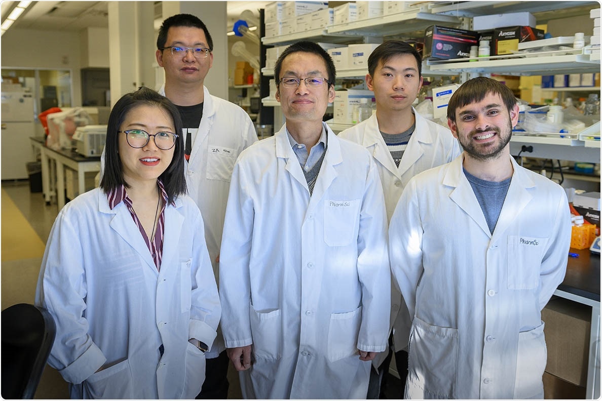
From left to right, study authors Yuening Liu, Peng Xia, Zhaokang Cheng, and Jingrui Chen and graduate student Joshua Gallo. Their work to uncover the mechanisms by which heart muscle cells die after heart attack treatment could someday lead to new treatment strategies to increase the survival and lifespan of heart attack victims. Image Credit: Cori Kogan, WSU Health Sciences Spokane.
According to this study, protein kinase A (PKA) plays a critical role in the necrosis of heart muscle cells. Necrosis is a major form of cell death that often takes place after reperfusion therapy—a medical treatment in which arteries are unblocked and blood flow is restored after a heart attack. The study has been published in the Journal of Biological Chemistry.
Our study has found that turning off a gene that controls this protein activity increased necrotic cell death and led to more heart injury and worse heart function following heart attack in a rodent model.”
Zhaokang Cheng, Study Author and Assistant Professor, College of Pharmacy and Pharmaceutical Sciences, Washington State University
Cheng continued, “With further research, this discovery could ultimately lead to the development of a small-molecule drug that could intervene in that pathway to limit or prevent heart muscle cell death after reperfusion therapy.”
A drug like that can help minimize heart injury and boost the survival and lifetime of heart attack patients, added Cheng. Each year, around 800,000 individuals in the United States experience a heart attack and this figure amounts to one heart attack taking place every 40 seconds.
Reperfusion therapy, in which mechanical means or clot-dissolving medications are used to unblock the clogged arteries, has traditionally been the most effective regimen for heart attacks. While this treatment considerably decreases heart damage, patients subjected to this regimen continue to experience certain damage, around half of which is actually caused by the therapy itself.
This is because when blood flow into oxygen-deprived heart tissues is rapidly restored, it causes a spike in free radicals. If this is left unchecked, the increased free radicals promote oxidative stress, which can destroy the heart muscle cells and cause heart damage as part of a condition called ischemia/reperfusion injury.
For a long time, investigators have regarded necrosis as an unavoidable, passive form of cell death, but new studies have indicated that some forms of necrosis occur in a highly controlled way that could be targeted for treatment. But scientists were not clear how this kind of cell death is actually controlled, and this aspect prompted Cheng to take a closer look.
Previously, Cheng and his research team had screened over 20,000 genes to search for ones that seemed to either promote or suppress necrotic cell death. They observed that PRKAR1A is one gene that required additional studies. This gene helps control PKA activity by encoding a protein called PKA regulatory subunit R1alpha.
Hence, the researchers performed a range of experiments to check out whether the R1alpha protein is capable of regulating necrotic cell death in a rodent model. The team observed that when the PRKAR1A gene is turned off, it increased cell death—both in mice and in cultured cells. PRKAR1A-gene-deficient mice also had worse heart function after a heart attack and more heart injury when compared to the wild-type mice.
Under normal conditions, the sudden spike of free radicals after heart attack therapy activates the heart to launch its own antioxidant defense system—the innate protective mechanism that helps maintain free radicals explained Cheng.
According to these findings, when the R1alpha protein is eliminated from the heart, the antioxidant defense system cannot be introduced as effectively as possible and this leads to more oxidative stress, which eventually causes cell death and heart damage.
Cheng explained that the R1alpha protein attaches to another form of a protein called PKA catalytic subunits to maintain the PKA activity. When the R1alpha protein is eliminated, the catalytic subunits are not regulated and the PKA activity thus increases, which according to Cheng is what inhibits the stimulation of the antioxidant defense system.
This indicates that the use of a small-molecule compound that preferentially suppresses PKA activity may block necrotic cell death and result in more improved treatment outcomes after heart attacks.
Although Cheng believes that the new study would ultimately enable the researchers to test this compound in an animal model, they are pursuing more studies to find out whether there are other mechanisms through which PKA controls necrotic cell death apart from the antioxidant defense system.
Source:
Journal reference:
Liu, Y., et al. (2021) Loss of PKA regulatory subunit 1α aggravates cardiomyocyte necrosis and myocardial ischemia/reperfusion injury. Journal of Biological Chemistry. doi.org/10.1016/j.jbc.2021.100850.