Reviewed by Danielle Ellis, B.Sc.Jun 3 2022
Professor Zi-Bing Jin is in charge of this research (Beijing Institute of Ophthalmology, Beijing Tongren Eye Center, Beijing Tongren Hospital, and Capital Medical University). He has spent the last decade working on retinal organoids, which can accurately mimic retinal development.
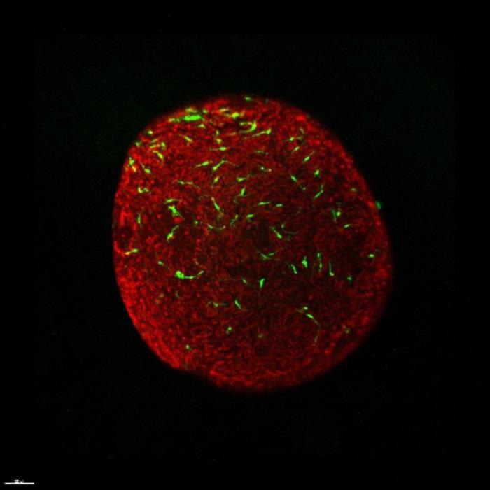
 Reconstructed 3D images showed that microglia were located underneath the photoreceptor cell layer. Green - microglia; Red - photoreceptor cells. Image Credit: ©Science China Press
Retinal organoids provide an excellent research tool to address fundamental questions, understand disease delineation and discover new drugs.”
Zi-Bing Jin, Professor, Beijing Institute of Ophthalmology
Microglia, the important neuro-immune cells that regulate the retina’s natural microenvironment, are missing entirely from the recently created retinal organoids, making them less representational of genuine retinas.
The researchers discovered macrophage and brain microglia differentiation and that there are techniques that are published earlier.
We try to generate retinal microglia in our lab because the macrophage and microglia are all from hematopoietic progenitors. Finally, we get it, a very simple and high-efficiency method to produce retinal microglia.”
Zi-Bing Jin, Professor, Beijing Institute of Ophthalmology
Researchers also noticed that differentiated microglia could identify damaged cells or cell debris from healthy cells and use phagocytosis to clean up the microenvironment. They can perform the same functions as microglia in vivo.
Scientists continually come up with new and fantastic ideas. The differentiated microglia were co-cultured with retinal organoids.
No one has ever done that before. It is awesome when I see the microglia migrate to the right position, proliferate and become retina-resident microglia in 3D retinal organoids.”
Zi-Bing Jin, Professor, Beijing Institute of Ophthalmology
These fresh and interesting findings show that the team can create “smart” microglia that can travel into retinal organoids and intentionally “find their way around.” They are functionally correct and correctly positioned. The retinal organoids are brought closer to the retinal organ.
The team presents a new framework of “integral retinal organs” for analyzing retinal microglial biology, retinal development, and retinal degenerative diseases, including a more efficient protocol for generating microglia from human pluripotent cells and confirmation that these microglia could be further distinguished into retina-resident microglia in a 3-dimensional retinal niche.
Source:
Journal reference:
Gao, M-L., et al. (2022) Functional microglia derived from human pluripotent stem cells empower retinal organ. Science China Life Sciences. doi.org/10.1007/s11427-021-2086-0.