Reviewed by Danielle Ellis, B.Sc.Oct 3 2022
The RIKEN discovery of a fluorescent protein produced from a Japanese jellyfish that maintains its brightness even under intense light could have significant positive effects on fluorescence imaging of biological samples.
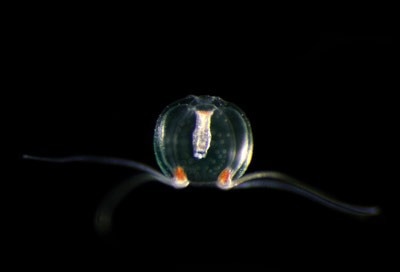
 RIKEN researchers have derived a photostable and bright green fluorescent protein from the Japanese jellyfish Cytaeis uchidae. Image Credit: © 2022 Kuroshio Biological Research Foundation
Proteins that emit green light when illuminated are effective instruments for capturing images of intricate cell structures. Such fluorescent proteins can be attached to target structures of interest, which light up when exposed to blue light.
To avoid interfering with normal biological functions, researchers want to utilize as little fluorescent protein as possible, yet doing so requires employing powerful light to get high-quality images.
The problem is that a fluorescent protein’s brightness rapidly decreases under intense light due to a process called photobleaching. The trade-off between brightness and photostability further complicates issues because enhancing one will almost always result in a decrease in the other.
Now, a fluorescent protein that defies this trade-off relationship has been developed by Atsushi Miyawaki of the RIKEN Center for Brain Science and his colleagues. It provides great brightness while being around 10 times more photostable than the best commercial fluorescent proteins.
The fluorescent protein, appropriately called StayGold, is made from a naturally occurring fluorescent protein that can be found in Cytaeis uchidae, a small jellyfish that can be found off the coast of Japan.
The discovery had a hint of coincidence to it.
We noticed that the fluorescent protein from the jellyfish was photostable but very dim. And I wasn’t optimistic about making the protein brighter while keeping that photostability, because I simply believed the tradeoff. However, to our surprise, we were able to increase both the protein’s photostability and its brightness. So could have our cake and eat it too.”
Atsushi Miyawaki, Team Leader, Cell Function Dynamics, RIKEN Center for Brain Science
With improved spatiotemporal resolution and length of observation, the researchers used StayGold to scan the endoplasmic reticulum network and mitochondria in cells. Additionally, they used it to visualize SARS-CoV-2, the virus that produces COVID-19, spike protein in infected cells.
The study has drawn a great deal of interest, as evidenced by the fact that over 44,000 people have accessed it since it was published in late April. The RIKEN BioResource Research Center, where researchers can try the protein, can provide it.
Miyawaki and his team plan to look into the mechanism underlying the fact that StayGold can both be brilliant and keep bright under illumination.
Source:
Journal reference:
Hirano, M., et al. (2022). A highly photostable and bright green fluorescent protein. Nature Biotechnology. doi.org/10.1038/s41587-022-01278-2