 By Pooja Toshniwal PahariaReviewed by Sarah KellyOct 23 2025
Swallowed like a capsule, this tetherless ‘pill that prints’ uses magnets and near-infrared light to precisely deposit living or cell-laden bioinks in the body, offering a glimpse of needle-free, surgery-free tissue repair.
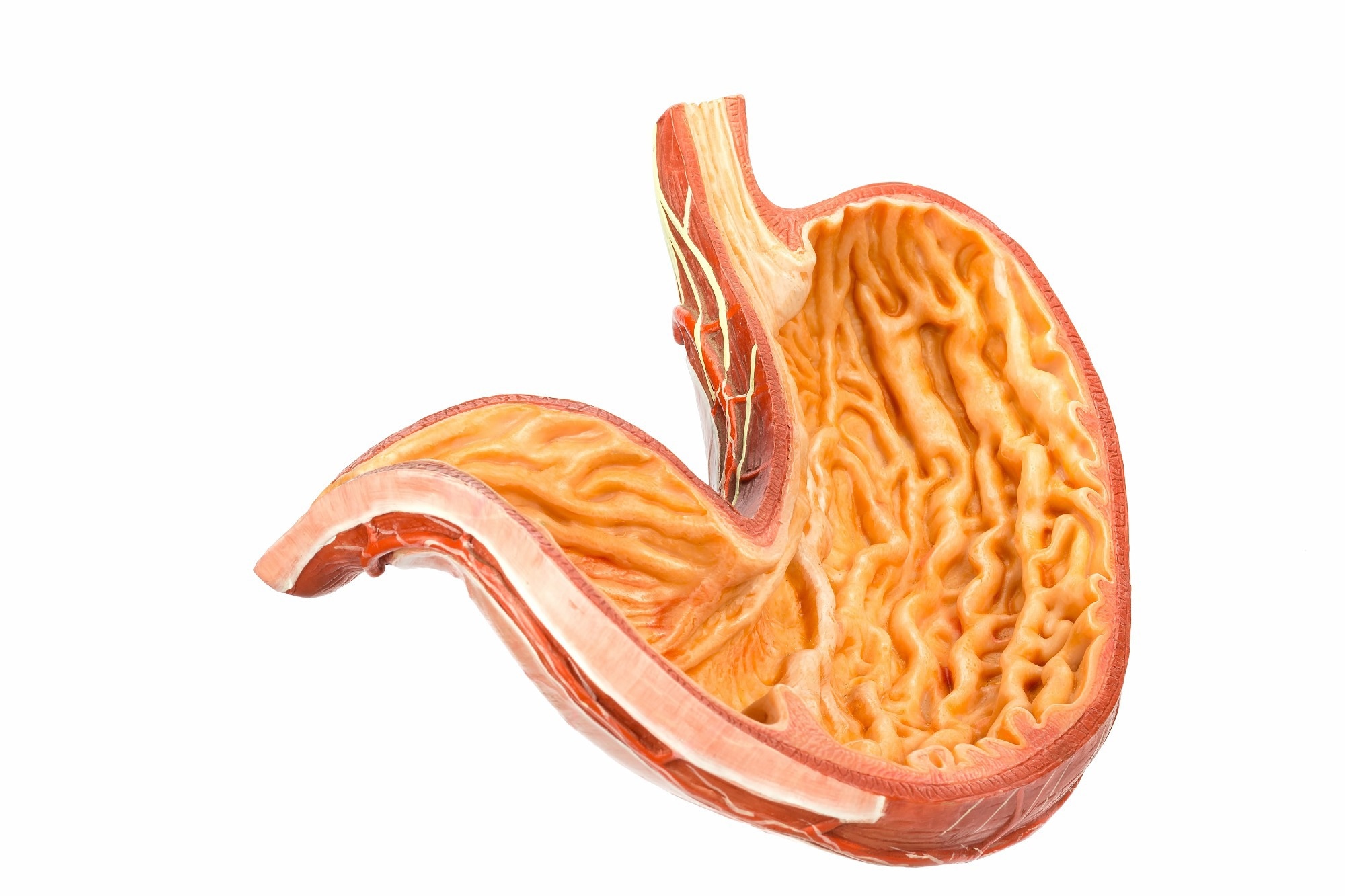
 Study: A Pill That Prints-An Ingestible Bioprinter for Non-Invasive Structured Bioink Deposition. Image credit: Ben Schonewille/Shutterstock.com
In a recent study in Advanced Sciences, researchers have introduced the first ingestible bioprinter that navigates the intestinal tract to deposit living bioinks onto internal tissues.
Despite advances in regenerative medicine, minimally invasive repair of internal tissues remains a critical challenge, particularly in gastrointestinal disorders such as ulcerative colitis and inflammatory bowel disease. Current therapies manage symptoms but cannot restore damaged tissue.
Conventional tissue engineering approaches, including injectable hydrogels and implanted scaffolds, require surgery, limiting their applicability in fragile or hard-to-reach GI regions.
While in-situ bioprinters and magnetically guided ingestible devices offer improved precision and reduced invasiveness, previous systems do not enable untethered, controlled deposition and patterning of bioinks directly onto internal tissue surfaces.
This work introduces a foundational demonstration of a miniaturized, tetherless in-situ bioprinter intended for preliminary feasibility rather than therapeutic evaluation.
About the Study
In this study, researchers developed and tested the Magnetic Endoluminal Deposition System (MEDS), a tetherless, ingestible bioprinter designed for non-intrusive tissue reconstruction within the gastrointestinal tract. The device integrates magnetic actuation and near-infrared (NIR) triggering to achieve wireless navigation, positioning, and bioink deposition without surgical access or onboard electronics.
The MEDS device comprises a biocompatible resin barrel with a 14 mm height and a six mm outer diameter. It contains a compression-spring plunger held by an NIR-responsive shape memory polymer (SMP) crossbeam
Upon NIR illumination, the SMP contracts, releasing the spring and extruding bioink through a gold-coated magnetic nozzle, which also serves as the effector magnet (EM) for steering. An actuator magnet (AM) mounted on a robotic arm guides MEDS through magnetic coupling, providing external control.
A unique magnetically modulated stick-slip (MMSS) locomotion mechanism enabled controlled movement along tissue surfaces. A novel ‘dispense-and-daub’ (DnD) bioprinting approach simplified printing, extruding the bioink at the target site and spreading it into defined patterns by manipulating the EM trajectory.
Barbed-ejector designs, validated through computational fluid dynamics, improved bioink coalescence and reduced topographical irregularities, enhancing print uniformity.
For validation, researchers first tested MEDS on ex vivo gastric tissue and silicone stomach phantoms to assess positioning accuracy, bioink extrusion, and lesion sealing.
They printed alginate-based bioinks crosslinked with calcium chloride, achieving stable adhesion and pattern fidelity under simulated gastric conditions. The device effectively sealed artificially created ulcers and hemorrhage models on ex vivo tissue.
Lastly, the team conducted in vivo feasibility studies in anesthetized rabbits. Under real-time fluoroscopic guidance, they orally introduced MEDS, magnetically guiding it to the stomach, and externally triggering bioink printing onto gastric tissue. After printing, they magnetically retrieved the device via the oral route, demonstrating a needle-free workflow, from ingestion and steering to tissue patterning and recovery.
Results
The study successfully demonstrated the design, control, and functional performance of the novel, tetherless MEDS system. Operated entirely through external magnetic and NIR control, MEDS enabled accurate bioink deposition and patterning in ex vivo and in vivo gastric environments.
NIR-triggered shape-memory beams, made from polylactic acid and melanin, showed over 90 % structural stability and 80 % functional recovery across all concentrations, confirming reliable spring actuation and extrusion through a 7.5 mm-thick section of muscle tissue ex vivo.
A cube with AM of 18 mm, at five mm/s, provided optimal actuation, allowing full six degrees of freedom and a hemispherical working volume of 5747 mm3 under ideal laboratory conditions. This configuration eliminated common tether-related issues such as bending damage and limited rotational freedom, achieving complete 360 ° yaw, pitch, and roll control.
The MEDS demonstrated robust hemostatic capability by sealing artificial hemorrhages using alginate bioink, forming leak-proof barriers validated by flow sensor data. The device’s DnD 2.5D printing approach enabled accurate patterning of circles, spirals, and polygons without needing synchronized extrusion. Three wt.% alginate provided optimal print fidelity, ensuring shape retention and biocompatibility.
Ex vivo tests on gastric tissue confirmed that print aspect ratio, circularity, and area deviation increased with substrate curvature, yet concave structures with aspect ratios less than 6.6 showed minimal impact on print fidelity.
Printed alginate constructs retained approximately 73 % of their mass under simulated gastric conditions, confirming stability for short-term applications, such as ulcer dressings or hemorrhage sealing. Confocal microscopy showed no surface damage from device movement, only surface smoothing.
Cell-laden bioinks containing human gastric fibroblasts maintained structural integrity and viability beyond 16 days, indicating potential for micro-bioreactor or regenerative applications. Preliminary in vivo studies in rabbits confirmed the practicability of fluoroscopy-assisted navigation, controlled extrusion, and patterned deposition on the stomach ceiling.
Conclusion
The MEDS system represents a groundbreaking advance in minimally invasive gastrointestinal therapies, enabling high-accuracy, tetherless bioink deposition for targeted repair of ulcers, hemorrhages, and other internal lesions. Integrating magnetic actuation with near-infrared control transforms internal tissue repair from extensive invasive procedures to pill-sized non-surgical bioprinting.
However, the researchers emphasized that these findings demonstrate technical feasibility only and that therapeutic efficacy and healing outcomes were not assessed in this study.
Future directions include extending the magnetic working range, implementing automated navigation, and refining bioink formulations to enhance cell viability and tissue integration, laying the foundation for next-generation, patient-friendly endoluminal regenerative therapies.
Journal Reference
Manoharan, S., & Subramanian, V. (2025). A Pill That Prints-An Ingestible Bioprinter for Non-Invasive Structured Bioink Deposition. Advanced Science, e12411. DOI:10.1002/advs.202512411
https://advanced.onlinelibrary.wiley.com/doi/10.1002/advs.202512411