Bruker's new Inverter Module for Ultima 2Pplus and Investigator Plus multiphoton microscopes facilitates inverted two-photon imaging while maintaining optical performance.
The Inverter Module rapidly and easily converts Bruker's upright multiphoton platforms into a versatile inverted configuration, enabling high-quality imaging of slides, dishes, organoids, and plated cells while retaining the original upright system's characteristics.
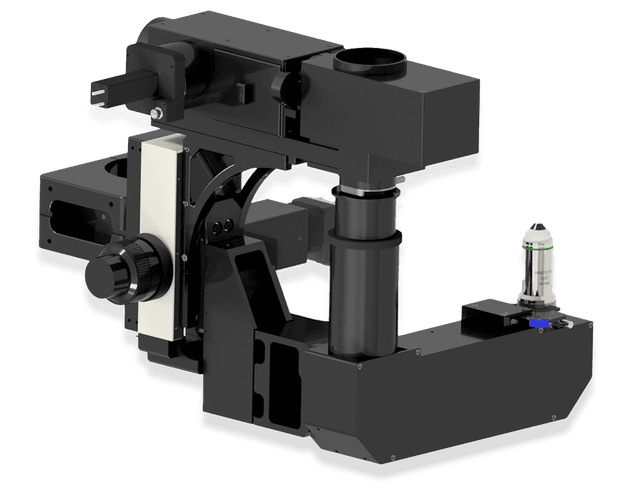
Bruker's Inverter Module for Ultima 2Pplus and Ultima Investigator. Image Credit: Bruker Nano Surfaces and Metrology
The Inverter Module was created to meet the demand for efficiently imaging samples that sink or stick to the bottom of dishes and plates. Using specialized relay optics and a mirror-based light-routing component, the module directs excitation and detection paths so that the objective lens faces upward, resulting in a genuine inverted arrangement.
The end result is a single device that can accommodate both upright and inverted imaging modalities, increasing experimental versatility and decreasing the requirement for separate microscopes.
Features
How the Inverter Module Extends Ultima System Capabilities
Inverted Configuration: When to Choose It | Fluorescence Microscopy Explained
Video Credit: Bruker Nano Surfaces and Metrology
True Upright‑to‑Inverted Conversion
Complete Optical Reorientation
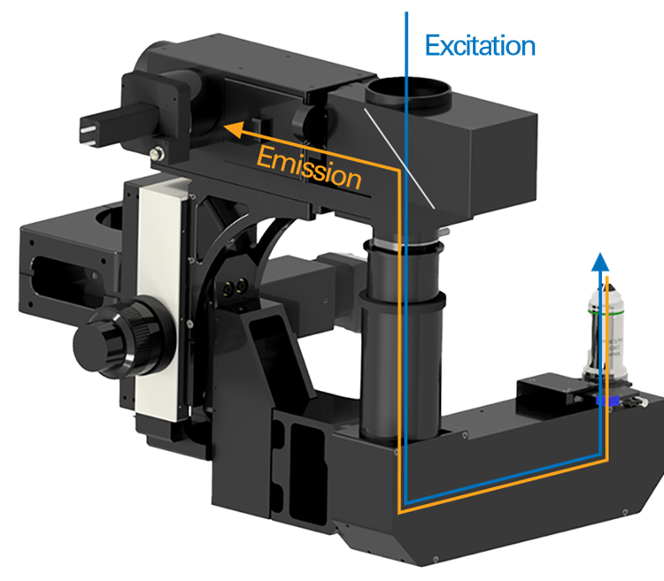
Bruker's Inverter Module for Ultima 2Pplus and Ultima Investigator. Image Credit: Bruker Nano Surfaces and Metrology
The Inverter Module completely reorients the optical geometry of the device, facilitating real upright-to-inverted conversions. The proprietary relay lens and mirror-based light-routing system redirects the excitation and detection routes so the objective points upward instead of downward, positioning the sample above the objective. This geometry is appropriate for slides, dishes, multi-well plates, and plated tissues.
Preserved Optical Performance
The Inverter Module was created to keep Bruker multiphoton systems running smoothly, avoiding the light loss and poor image quality that come with workarounds and incomplete changes. Its innovative optics and mechanics provide complete signal throughput, optical alignment, and image integrity, allowing for inverted imaging without sacrificing quality.
No Second System Required
The Inverter Module transforms a single system into a dual-purpose platform capable of upright and inverted multiphoton imaging. The same tool can image dish-based experiments, organoids, plated cells, and tissue samples, enabling in vivo and ex vivo neuroscience research. This removes the requirement for a separate inverted microscope.
Purpose‑Built for Safe, Simple, Real‑World Operation
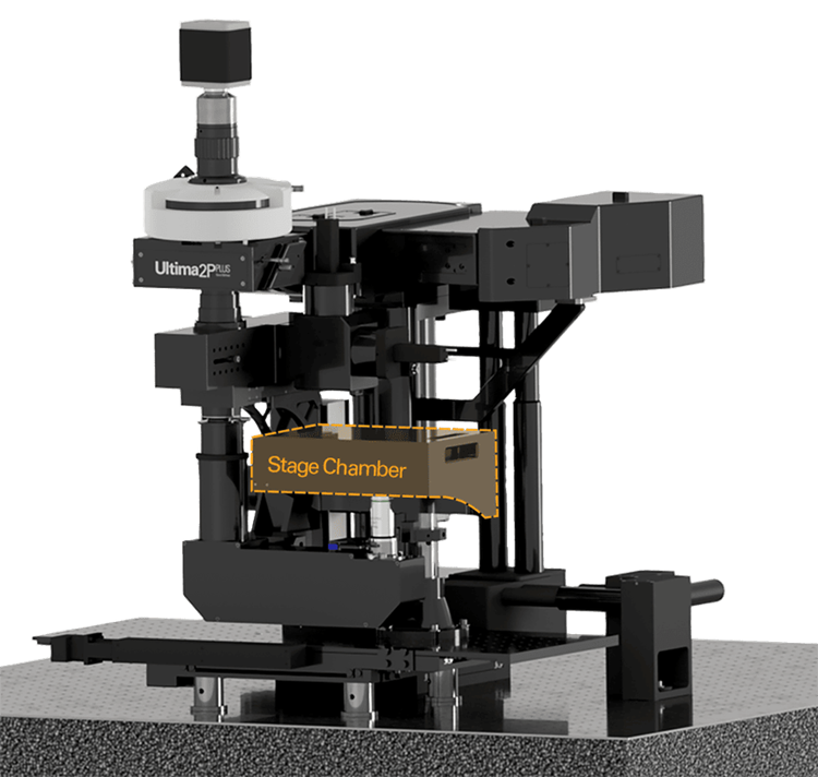
Imaging stage chamber compatible with slides, dishes, and well plates. Image Credit: Bruker Nano Surfaces and Metrology
Seamless On-Site Integration
The Inverter Module was developed for direct integration with Bruker multiphoton systems and includes full on-site installation. Installation may sometimes be completed in a single day, reducing research disruption and allowing labs to expand their capabilities swiftly.
Intuitive Dual‑Mode Operation
Once implemented, users can simply switch between upright multiphoton imaging and inverted operations on their own. Researchers can use Bruker's standard XY stage to access inverted formats such as slides, dishes, tissues, and multi-well plates while maintaining their familiar motion control and sample-handling procedures.
Safe for Inverted Configurations
To operate safely in an inverted geometry, a light-tight, safety-interlocked chamber prohibits exposure to upward-facing laser beams. The laser can only be used while the enclosure is closed, assuring safety in both individual labs and shared facilities.
Applications
Supporting Advanced Imaging across Key Research Areas
Neuroscience
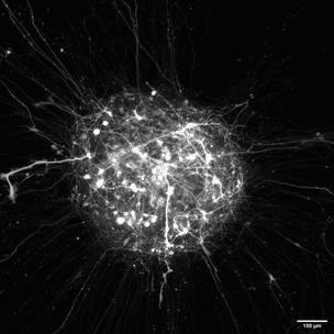
Image Credit: Bruker Nano Surfaces and Metrology
The Inverter Module enables dish-based neuroscience operations, such as organoid validation before implantation and high-quality imaging of brain slices made in dishes or chambers.
Organoid Research
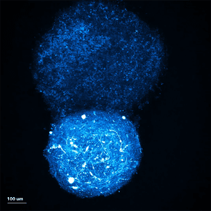
Image Credit: Bruker Nano Surfaces and Metrology
The system allows for two-photon imaging of organoids grown in plates and dishes, as well as multi-well screening with XY-stage scanning for high-throughput research.
Cancer Biology
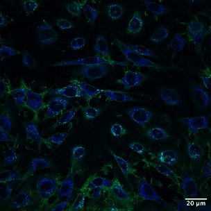
Image Credit: Bruker Nano Surfaces and Metrology
The Inverter Module allows researchers to capture GFP and autofluorescence signals while imaging plated tumor samples and fresh tissue slices, allowing for in-depth examination of tumor metabolism and structure.
Immunology and Cell Biology
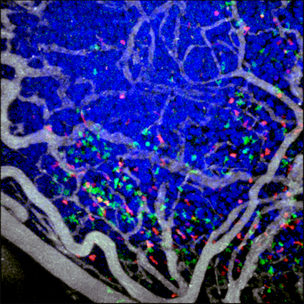
Image Credit: Bruker Nano Surfaces and Metrology
The Inverter Module enables high-resolution imaging of plated cells and tissues, and is compatible with multiphoton imaging in conventional dish and well-plate formats for cell-based experiments.
Use Cases
Examples from Current Research
Imaging of Neural Circuits in Live Spheroids
The Inverter Module facilitates organoid workflows in dish-based and in vivo situations, as illustrated by the following two examples.
In Figure 1, organoids grown and plated on plates were scanned using the Inverter Module, enabling the lab to perform high-quality two-photon imaging in an inverted configuration designed for dish-based materials.
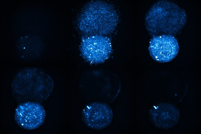
Figure 1. Fusion of healthy and diseased IPSC neuro-spheroid labeled with JF552. Sample courtesy of Trish Hoang, Promega. Image Credit: Bruker Nano Surfaces and Metrology
Figure 2 shows neural circuits built in a live spheroid, with organoids generated and confirmed ex vivo in an inverted form before implantation for upright in vivo multiphoton imaging. The inverter module streamlines the workflow from dish-based validation to in vivo imaging, facilitating investigations on transplantation, integration, and regeneration.
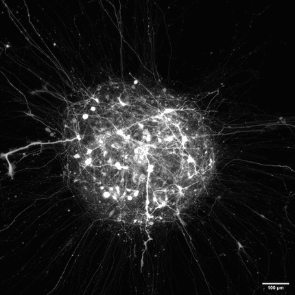
Image 2. Neural circuits developed in a live spheroid imaged with the Ultima 2Pplus in an inverted configuration. Sample courtesy of Trish Hoang, Promega. Image Credit: Bruker Nano Surfaces and Metrology
Inverted Imaging of Cancer Cells Reveals Metabolic Insights
Researchers used the Inverter Module to image plated cancer tissue samples, which included multicolor and autofluorescence imaging. Large tissue regions could be obtained via XY-stage scanning and assembled into high-resolution montages to aid metabolic and structural research.
Figure 3: The Inverter Module was used to examine fresh, human-derived cancer tissue, capturing GFP and native autofluorescence signals in an inverted configuration. The technique resulted in high-quality two-photon images of actual tissue samples in conventional dish formats.
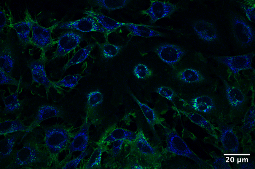
Figure 3. Live melanoma cells expressing membrane-bound GFP. Blue Channel: excitation 740, emission 460/50 (mainly autofluorescence). Green Channel: excitation 860 nm, emission 525/50 (GFP). Data Courtesy of Matt Lindley, Irene Georgakoudi Lab, Dartmouth College. Samples provided by Tyler Curiel Lab, Dartmouth College. Image Credit: Bruker Nano Surfaces and Metrology
Figure 4: A stitched-field acquisition was used to image a bigger sample of cancer tissue. The XY stage was used to gather several neighboring fields of view and combine them into a continuous multicolored picture. Large-area, plate-based tissue imaging that is appropriate for analyzing tumor shape and metabolism was made possible by this method.
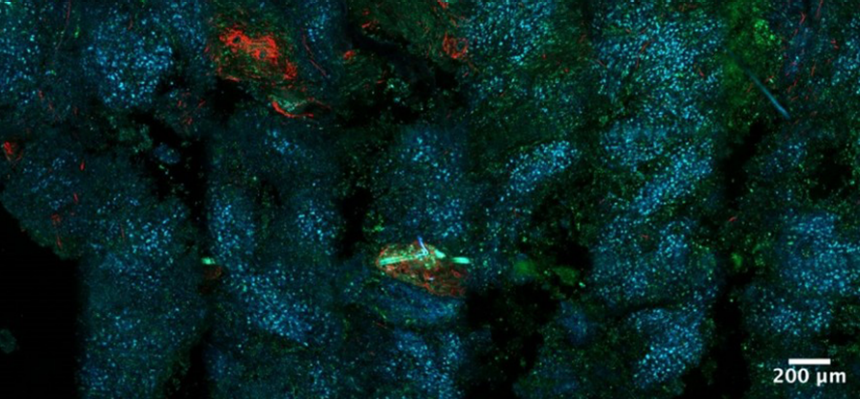
Figure 4. Label-free tile-scan of a fresh melanoma tumor from a mouse model. Data courtesy of Matt Lindley, Irene Georgakoudi Lab, Dartmouth College. Samples provided by Tyler Curiel Lab, Dartmouth College. Image Credit: Bruker Nano Surfaces and Metrology
Compatible Multiphoton Imaging Systems
Ultima 2Pplus
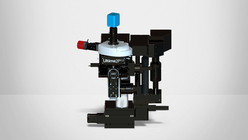
Image Credit: Bruker Nano Surfaces and Metrology
A complete all-optical multiphoton workstation for imaging and optical manipulations.
Ultima Investigator Plus
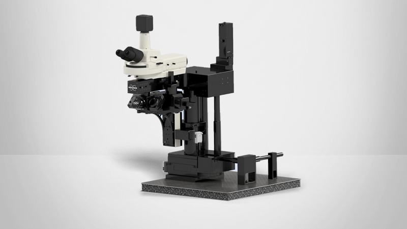
Image Credit: Bruker Nano Surfaces and Metrology
A large field-of-view, easily accessible multiphoton microscope that expands to accommodate the study.