The biological intracellular microenvironment is diverse, consisting of several cell compartments and intracellular components. The development of micro/nanoprobes for subcellular assessment is one of the most important components in properly characterizing the physiological activity of live cells.

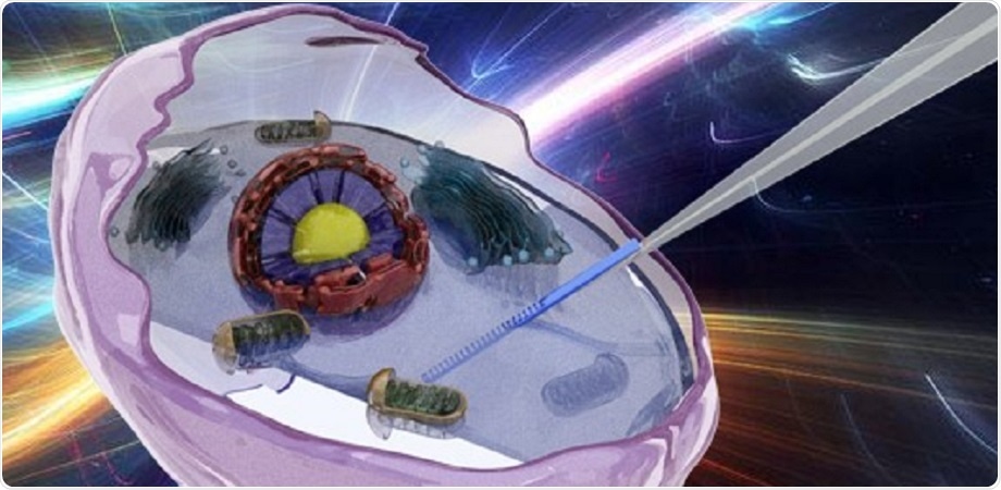
Integrated fiber probe for label-free detection inside living cells. Image Credit: Li et al.
Dyes and quantum dot-doped photoelectric materials are often used as sensors or calibration objects in current nanoprobe methods, which are paired with far-field super-resolution optical technology. However, these methods lack a potential circuit that can track both interior and exterior interactions between photoelectric signals and molecules. Background fluorescence interference and bleaching also influence the accuracy of long-term measurement outcomes.
A micro/nano-sized fiber is well adapted to the nanoscale and sub-milliscale of normal cells as a platform for intracellular sensing and control, allowing lossless transmission of far-near-field optical signals. In order to maintain the survival of the cells in vitro, the incision needed for probe insertion must be less than 1 μm. Given this limitation, the optimal micro/nanofiber probe’s sensing functional area must be constrained.
With its low refractive index, it is challenging to see a sensor tip on the order of micrometers or even nanometers inside the framework of a silica optical fiber (RI). External materials and structures are introduced and integrated into fiber devices to achieve a fiber probe with a complete spectrum function module for long-term nonfluorescent detection while reducing the size of the device.
Nanjing University researchers recently constructed a multifunctional, biocompatible, portable, and reusable microfiber probe based on a zinc oxide (ZnO) nanograting-integrated microfiber, as mentioned in the study published in the journal Advanced Photonics. It functions as an RI sensor for real-time monitoring of cellular molecules and live, label-free sensing of intracellular RI distribution.
A ZnO nanowire with gratings etched on the front end and a fiber taper probe make up the device. The device’s sensing area is roughly 800 nm × 6 μm, which is significantly smaller than that of standard fiber gratings.
This innovative nano-optical functional module combines signal transmission, detection, and collection, considerably enhancing operability and sensitivity. For long-term single-cell detection, nanowire gratings, rather than fluorescent particles, provide a steadier and dependable performance.
The researchers used a single living HeLa cell to show the nanowire probe’s activity. HeLa cells are human cancer cells named after Henrietta Lacks, a cancer patient from whom the cell line was extracted. Owing to the device’s sensitivity to RI, the researchers were able to monitor changes in cell morphology and the intracellular microenvironment during apoptosis—a stage of cell growth and maturation.
Quantitative identification and study of the refractive indices’ natural sources in single live cells during apoptosis can help in the advancement of knowledge about cell life occurrences and disorders.
Using a label-free nano-optical device in live HeLa cells, this study set a precedent for real-time, in situ detecting and tracking of early cellular apoptosis. It will not only provide a scientific understanding of cell life events, but it will also extend the scope of fiber nanosensor applications.
Simultaneously, introducing functional nanomodules into single cells reduces limitations to getting information on the interactions between interior and exterior cell media, paving the door for novel approaches to combine the benefits of fiber optics with cell biology.
The evolution of cell physiological processes may be further researched in the future, and real-time monitoring of temperature and substance concentration in cells may be accomplished through the creation of diverse structures.
Source:
Journal reference:
Li, D., et al. (2022) Label-free fiber nanograting sensor for real-time in situ early monitoring of cellular apoptosis. Advanced Photonics. doi.org/10.1117/1.AP.4.1.016001