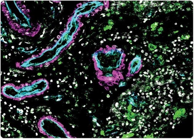
Blood vessels in this microscope image of an AVM (cyan and blue) contain patches of immune cells (green) that destabilize the vasculature, potentially leading to stroke. Image Credit: Jayden Ross and Ethan Winkler.
The atlas identifies more than 40 previously unknown cell types, including a population of immune cells whose connection with vascular cells in the brain contributes to hemorrhagic stroke bleeding. This deadly kind of stroke accounts for 10–15% of all strokes in the United States, with the majority of victims being young adults. Around half of Hemorrhagic strokes are deadly.
The research will serve as a platform for future study on brain vasculature throughout the world, according to the researchers.
This research gives us the map and the list of targets to start developing new therapies that could change the way we treat a lot of cerebrovascular diseases.”
Ethan Winkler MD, PhD, Study Lead Author, Neurosurgeon and Research Associate, Weill Institute for Neurosciences, University of California San Francisco
This study was published on January 27th, 2022, in the journal Science.
Tangles in the brain’s vasculature
The researchers examined cells in arteriovenous malformations, or AVMs, which are tangles of poorly formed arteries in the brain that are often the source of a hemorrhagic stroke. The team was led by Adib Abla MD, associate professor of Neurological Surgery, and Daniel Lim MD, PhD, professor of Neurological Surgery, both being members of the UCSF Weill Institute for Neuroscience, with Tomasz Nowakowski, PhD.
They compared the AVMs to samples of normal brain vasculature collected from five volunteers who were having epilepsy surgery.
UCSF is a top national center for brain AVM surgery and treatment, having been named #1 in neurosurgery by U.S. News and World Report.
Some of the 44 samples of AVM tissue obtained during delicate surgeries done by Abla, Chief of Neurological Surgery, were taken from the patient’s brain while they are still intact, while others were only removed after they began to bleed. The three types of tissue—normal, intact AVMs, and AVMs that bled—allowed the researchers to gain a better understanding of the distinctions between how the cells behave normally and in different states of illness.
The scientists employed single-cell mRNA sequencing on more than 180,000 cells in partnership with the Cerebrovascular Research Center to discover which genes were expressed in different samples and linked gene expression with cell location. Chang Kim, a UCSF bioinformatics graduate student and research co-lead author, created computer studies that analyzed gene expression in normal and diseased cells.
An immune cell surprises
The findings revealed not just a wide range of novel cell types, but also a population of immune cells that appear to communicate with smooth muscle cells in diseased arteries, weakening them and causing a stroke. Malformations like AVMs are thought to trigger the immune system, according to scientists.
“Without this study, we wouldn’t be able to pinpoint this very specific population of cells in the blood that might be the key drivers of disease progression,” added Nowakowski.
Nowakowski continued by stating that identifying these particular immune cells has completely changed how researchers think about treating vascular disease. It may be possible to minimize stroke risk by manipulating the immune system if the cells circulate in the bloodstream.
“This opens up huge therapeutic potential,” explained Nowakowski.
That capacity extends past stroke. The map can assist in checking out any neurovascular disease, consisting of one of the maximum common disorders—dementia.
Many forms of dementia, including Alzheimer’s, appear to have a vascular underpinning. We need an atlas like this to better understand how changes in the vasculature can contribute to the loss of cognition and memory.”
Daniel Lim MD, PhD, Professor, Neurological Surgery, Weill Institute for Neurosciences, University of California San Francisco
Daniel Lim states, “This work was a really a beautiful collaboration between surgeon-scientists and molecular biologists, occurring in a place with incredible access to clinical specimens. It’s what makes the Weill Institute of Neuroscience at UCSF so special.”
Although many establishments do not have entry to all of those important resources, they will have access to the dataset from this study, Lim added. Nowakowski considers that these facts will permit researchers across the globe to do cheaper analyses on massive numbers of patients, which will give us a better understanding of how vascular diseases operate.
“Understanding cerebrovascular disease at the cellular and molecular level will take the work of many researchers into new directions,” Lim remarked.
A “periodic table of cell types”
The research contributes to the Human Cell Atlas, a global project to develop cell reference maps for the whole body.
These atlases are described by Nowakowski as a “periodic table of cell types.” Just as the chemical periodic table arranges components into a structure that allows scientists to infer correlations between them depending on where they occur in the table, human cell atlases disclose the locations of cells in the body and the consequent interactions between them.
While there is a lot of effort being taken to create these atlases for various organs and tissues all around the world, many of them just map the geographic locations of cells. This study takes it a step further by comparing normal and unusual cells, offering incredibly refined drug development assistance.
Our study really demonstrates how a cell atlas can be utilized. With our ‘periodic table’ as a reference, we can start asking which cells might go wrong in disease and very precisely target those cells for therapy.”
Tomasz Nowakowski PhD, Department of Neurological Surgery, University of California San Francisco
Source:
Journal reference:
Winkler, E. A., et al. (2022) A single-cell atlas of the normal and malformed human brain vasculature. Science. doi.org/10.1126/science.abi7377.