Reviewed by Danielle Ellis, B.Sc.Sep 14 2022
Researchers at the National Eye Institute (NEI) have demonstrated for the first time how cells across various tissue layers in the eye are impacted in individuals with choroideremia, a rare genetic disorder that causes blindness.
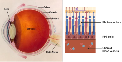
 On the left: diagram of the eye. On the right: layers within the retina and the adjacent choroid. Image Credit: The National Eye Institute.
This was accomplished by combining conventional eye imaging methods with adaptive optics, a technology that improves imaging resolution. Their research was published in Communications Biology and was supported by the NEI Intramural Research Program. NEI is part of the National Institutes of Health.
Indocyanine green dye and adaptive optics were used by Johnny Tam, Ph.D., head of the NEI Clinical and Translational Imaging Unit, to examine live retinal cells, including light-sensing photoreceptors, retinal pigment epithelium (RPE), and choroidal blood vessels.

Image Credit: HQuality/Shutterstock.com
The amount to which choroideremia damages these tissues was clearly visible to his team, and this knowledge might be used to develop efficient treatments for this and other disorders. The RPE, a layer of pigmented cells in the retina, is crucial for the maintenance and survival of photoreceptors.
Due to the gene’s location on the X chromosome, men are more likely than women to develop choroideremia. Males typically experience more severe symptoms due to the fact that they only have one copy of the X chromosome, whereas females, who have two copies of the X chromosome, typically experience milder symptoms due to the presence of one functional copy of the gene on the other X chromosome.
One major finding of our study was that the RPE cells are dramatically enlarged in males and females with choroideremia, We were surprised to see many cells enlarged by as much as five-fold.”
Johnny Tam, Head, Clinical and Translational Imaging Unit, National Eye Institute
RPE cells in the female study participants were both larger and appeared to be in better health. According to Tam, this may help to explain why women with choroideremia experience milder symptoms. Both male and female participants in the study had less impact on the photoreceptor and blood vessel layers, indicating that RPE disruption is a significant factor in choroideremia.
Regular diagnostic testing in eye clinics does not include Tam’s adaptive optics. However, his team discovered that enlarged RPE cells may be identified even when using just an indocyanine green dye and a scanning laser ophthalmoscope that is commercially available.
It’s not obvious at first, but using an existing tool in the clinic, we can monitor and track the cellular status of the RPE layer. This could prove valuable in identifying which patients would benefit the most from therapeutic interventions.”
Johnny Tam, Head, Clinical and Translational Imaging Unit, National Eye Institute
Source:
Journal reference:
Aguilera, N., et al. (2022) Widespread subclinical cellular changes revealed across a neural-epithelial-vascular complex in choroideremia using adaptive optics. Communications Biology. doi.org/10.1038/s42003-022-03842-7.