In the third volume of Psychoradiology, published in 2023, a group of researchers from The University of Hong Kong and The University of Electronic Science and Technology of China has definitively pinpointed the right inferior frontal gyrus (rIFG) as a pivotal input and causal regulator within the subcortical response inhibition nodes.
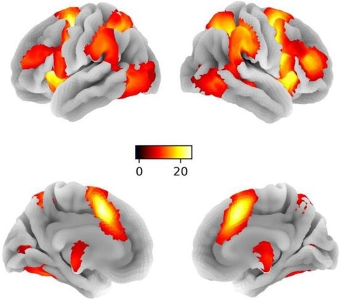
 Brain activation maps for general response inhibition on whole brain level (contrast: NoGo > Go; P < 0.05 FWE, peak level). L, left; R, right. The color bar represents the t-values of the BOLD signal and reflects the significance level of the contrast. Image Credit: Psychoradiology.
This inhibitory control circuit, which leans towards the right hemisphere, is characterized by substantial intrinsic connectivity, emphasizing the critical role of the rIFG in coordinating top-down cortical-subcortical control. This underscores the intricate dynamics of brain function in the context of response inhibition.
In their comprehensive study, the researchers utilized dynamic causal modeling (DCM-PEB) and functional magnetic resonance imaging (fMRI) with a sizable sample size (n = 250) to investigate inhibitory circuits in the brain. Their focus was particularly on the right inferior frontal gyrus (rIFG), caudate nucleus (rCau), globus pallidum (rGP), and thalamus (rThal).
Treating the brain as a nonlinear dynamical system, this approach allowed the estimation of directed causal influences among these nodes, taking into account task demands and biological variables.
The findings unveiled robust intrinsic connectivity within this neural circuit, with response inhibition notably strengthening causal projections from the rIFG to both rCau and rThal. This amplifies the regulatory role of the rIFG during such tasks.
The study also highlighted the significant impact of sex and performance metrics on the functional architecture of the circuit. For instance, women exhibited increased self-inhibition in the rThal and reduced modulation to the GP.
Meanwhile, better inhibitory performance correlated with more robust communication from the rThal to the rIFG. Notably, these communication patterns did not mirror in a left-lateralized model, emphasizing a hemispheric asymmetry.
The research suggests that different brain processes may mediate similar behavioral performances in response inhibition across genders, particularly in thalamic loops. Higher response inhibition accuracy was associated with stronger information flow from the rThal to the rIFG.
These insights into the brain's inhibitory control mechanisms hold significant implications for understanding various mental and neurological disorders characterized by response inhibition deficits.
The findings of the study could pave the way for the development of targeted neuromodulation strategies and personalized interventions to address these deficits, ultimately enhancing the treatment and management of such conditions.
Source:
Journal reference:
Zhuang, Q., et al. (2023) The right inferior frontal gyrus as pivotal node and effective regulator of the basal ganglia-thalamocortical response inhibition circuit. Psychoradiology. doi.org/10.1093/psyrad/kkad016.