 By Pooja Toshniwal PahariaReviewed by Lauren HardakerJul 25 2025
In a recent study published in Advanced Science, researchers developed a novel approach using biomimetic MXene-ECM composite scaffolds to enhance the delivery of therapeutic electrical stimulation (ES) to neurons. The scaffolds, which are three-dimensionally (3D) printed and incorporated with MXene-PCL micro-meshes in a hyaluronic acid (HA)-based extracellular matrix (ECM), effectively promote neurite outgrowth and neuronal differentiation in a spatially controlled manner.
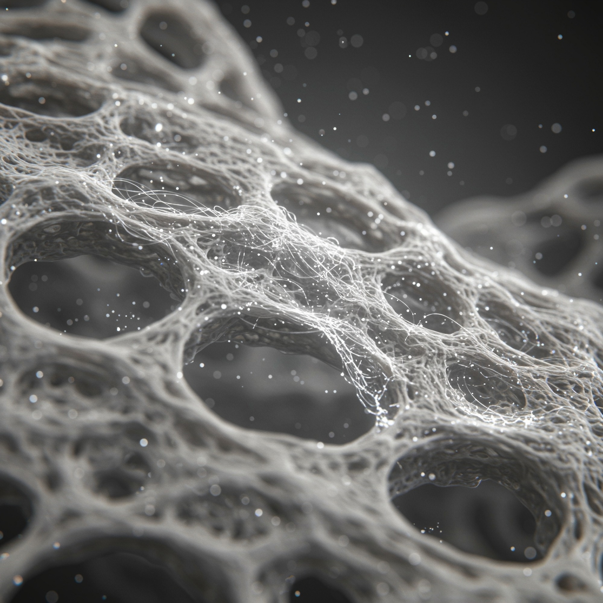
 Image credit: Shutterstock AI/Shutterstock.com
The study found that the effects of ES varied by micro-mesh design, with medium-density scaffolds yielding the best average neurite outgrowth for neuronal cell lines. In contrast, high-density scaffolds supported the greatest axonal extension in stem cell neurosphere models. This multi-pronged approach can treat complex neurotrauma injuries like spinal cord and traumatic brain injuries, promoting specific repair-critical effects like increased axonal length and density.
Neurotrauma, a condition causing severe cognitive, motor, and sensory impairments, can lead to paralysis and disability. The complex nature of neurotrauma, including inflammation, scarring, and poor neuronal regrowth, makes effective treatments challenging.
Electrical stimulation has potential as a therapeutic tool, promoting axonal growth, enhancing neuronal plasticity, supporting function, and activating key cell signaling pathways, including the mammalian target of rapamycin (mTOR); this study did not directly examine mTOR activation. However, traditional methods of ES delivery often involve metallic electrodes, which have limitations such as compliance mismatch, poor neurocompatibility, slow degradation, and release of toxic ions. Biomaterial-based strategies offer a solution by providing an environment that supports axonal growth and enables local therapeutic delivery.
About the Study
In the present study, researchers engineered a multifunctional tissue-engineered scaffold through 3D printing to increase the neuro-reparative effects of therapeutic ES.
The team integrated Ti₃C₂Tₓ MXene nanosheets into an ECM-based scaffold to build electroconductive scaffolds that could enhance ES delivery to ingrowing neurons. To do so, they synthesized MXene nanosheets from Al-rich Ti3AlC2 MAX-phase powder. A melt-electrowriting process printed highly organized polycaprolactone (PCL) micro-scale meshes comprising varied fiber spacings (low-, medium-, and high-densities of 500, 750, and 1000 µm, respectively). The process enabled spatially controlled MXene distribution. The PCL mesh design involved rectilinear patterns with layers swapped perpendicularly to create a box-like architecture.
The researchers functionalized the meshwork with MXenes to provide tunable electroconductive properties. They embedded the meshes in a neurotrophic, biomimetic, immunomodulatory HA-based ECM comprising fibronectin and collagen type IV. The design created a soft, growth-supportive scaffold simulating the anatomical features of nervous tissues. While the ECM-only phase showed a Young’s modulus of ~0.6 kPa (similar to brain tissue), the composite scaffolds ranged from ~1.3 kPa (low density) to ~3.25 kPa (high density), balancing softness with added mechanical reinforcement.
To assess biocompatibility, the team seeded the PCL films with SH-SY5Y human neuronal cells, murine IMG microglial cells, and human-derived astrocytes. The cells underwent ES (200 mV/mm, 12 Hz, 50 ms pulse for six days). Subsequently, the researchers evaluated cellular viability, metabolic activity, and neurite outgrowth.
Scanning electron microscopy (SEM) analyzed MXene film and scaffold structures, whereas energy-dispersive X-rays (EDX) determined the MXene composition. Atomic Force Microscopy (AFM) enabled surface topography analysis. Cyclic Voltammetry (CV) analysis investigated electrochemical properties, and uniaxial compression tests determined stress-strain behavior.
To examine the effects in multicellular backgrounds, the team subjected neurospheres derived from mice's olfactory bulbs to ES therapy for seven days. Immunohistochemical labeling followed by confocal microscopy revealed cellular differentiation and axon outgrowth from the neurospheres. Enzyme-linked immunosorbent assays measured the release of cytokines, interleukin-1β (IL-1β), and tumor necrosis factor-alpha (TNF-α), from activated cells.
Results
The micro-meshes, functionalized with MXenes, showed highly tunable electroconductive properties ranging from 0.08 to 18.87 S/m. The tunability allows precise control over electrical stimulation delivery, a significant advantage for optimizing ES therapeutic outcomes. For SH-SY5Y neurons, medium-density scaffolds significantly improved average neurite outgrowth (≈365 µm per cell) and βIII-tubulin expression compared to both low- and high-density designs, despite high-density scaffolds achieving the greatest bulk conductivity.
Optimizing the micro-mesh design can significantly influence neuronal responses to ES, with different density designs favoring specific outcomes for different cell models. This study showed that medium-density scaffolds optimized neurite outgrowth from SH-SY5Y cells. At the same time, high-density designs maximized axonal extension in neurospheres, demonstrating that scaffold architecture, not just conductivity, drives therapeutic outcomes.
The scaffold's biomimetic and mechanical properties closely match those of native brain tissues at the ECM-only phase (Young's modulus of 0.6 kPa). In comparison, the full composite scaffolds range from ~1.3–3.25 kPa depending on mesh density, and support primary neural cells such as neurons, microglia, and astrocytes without inducing significant cytotoxicity or inflammation. The biocompatibility of the MXene/PCL surfaces indicates that MXene coatings are suitable for implantation in neural tissues. Importantly, the MXene scaffolds showed reduced rather than absent signs of astrocyte reactivity as astrocytes on MXene/PCL substrates showed significantly decreased GFAP expression (P <0.05) compared to PCL controls, suggesting reduced, but not absent, reactivity.
The spatial organization of the electroconductive MXene micro-meshes within the neurotrophic ECM-based scaffold significantly enhanced the therapeutic effects of ES. Specifically, stimulated MXene-ECM samples showed significantly longer maximum axon length (108.5 ± 6.1 µm) compared to non-stimulated controls (74.3 ± 5.8 µm). However, the average neurite length did not differ significantly from that of PCL controls under the same ES conditions.
In the multicellular model using murine olfactory neurospheres, stimulation on high-density MXene-ECM scaffolds for seven days significantly increased axonal extension and neuronal cell differentiation compared to the low-density scaffolds and MXene-free controls. The high-density scaffolds demonstrated the highest axonal extension of 203.6 µm. Overall, MXene content was very low (<0.5% v/v), maintaining scaffold softness while providing electroconductivity, though bulk conductivity decreased by ~50% over seven days due to film disruption (not MXene degradation).
Conclusions
Based on the findings, the 3D-printed MXene-ECM multifunctional scaffolds show promise for neurotrauma repair, enhancing repair-critical responses to electric stimulation therapy. The study demonstrates that integrating electroconductive materials within a neurotrophic scaffold, combined with precise stimulation control, can significantly improve neural regeneration, paving the way for advanced neurotrauma treatments.
The results also highlight that scaffold design must be tailored, with medium density for optimal neurite outgrowth in neuronal cells and high density for maximal axonal extension in stem cell neurospheres, rather than relying solely on maximizing conductivity. Future studies could investigate the potential benefits of integrating the MXene-ECM composite scaffolds with existing neurotrauma therapies or in vivo brain or spinal cord stimulation.
Download your PDF copy now!
Journal Reference
Ian Woods et al. (2025). 3D-Printing of Electroconductive MXene-Based Micro-Meshes in a Biomimetic Hyaluronic Acid-Based Scaffold Directs and Enhances Electrical Stimulation for Neural Repair Applications. Adv. Sci., e03454, DOI: 10.1002/advs.202503454 https://advanced.onlinelibrary.wiley.com/doi/10.1002/advs.202503454